Blog
A short overview of the history of pathology: origins, early days, and the transition to novel technologies
By Anna Correas Grifoll
16 May, 2024
Throughout history, pathology has remained an integral component of medicine, tracing its origins to ancient healers recognizing the importance of understanding disease processes for effective treatments. What began as speculative observations has since evolved into a complex specialty characterized by precise assessments, predictive diagnosis, and advanced imaging modalities. This transformation has been driven by technological advances that have revolutionized this field time and time again.
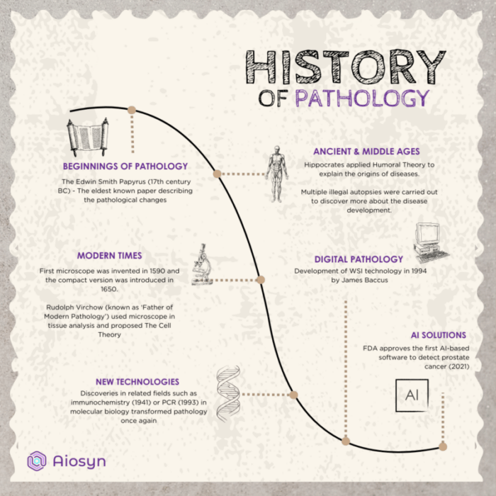
Ancient healers understood how important it is to map the disease’s development, describe the abnormal structures found in patients, and look for similarities in prevalent conditions. However, due to their constraints and lack of expertise, this was mostly limited to empirical assessments that often have a speculative nature. One of the first medical documents that can be traced back to the beginnings of pathology is the Edwin Smith Papyrus, which dates back to the 17th century BC and is written entirely in hieroglyphs [1]. It depicted skin ulcerations in patients; however, it was impossible to draw any causal relationships or map the development of the condition.
Hippocrates, often recognized as the father of medicine, was the first physician to propose the underlying mechanisms behind medical conditions in the form of the Humoral Theory [1]. In the 4th century BC, he theorized that humans consist of four vital fluids: blood, phlegm, yellow bile, and black bile. Hippocrates believed that once there was a significant excess or deficiency of any of those, the individual would fall ill. This theory was prevalent until it was disproved in the 17th century.
The Middle Ages brought the next major developments, as, although illegal, former physicians and healers performed autopsies and disseminations to better understand the nature of different conditions and the drivers behind them. Antonio Beniveni (1443‐1502) developed ‘De abditis Nonnullis ac Mirandis Morborum et Sanationum Causius’ [1], the first publication in which pathology findings appear separately and in a systematic way, with over 100 descriptions of cases he encountered during his research. This is often referred to as the time when pathology became a specialty on its own rather than just a part of medicine. Throughout the Middle Ages and Renaissance, physicians were systematically describing new cases and publishing their findings and theories, slowly straying away from Hippocrates’ Humoral Theory.
The first technological advance that elevated the field of pathology to a new level was the invention of microscopes. Zacharias Janssen invented the first microscope in 1590, and then Robert Hooke made it available in compact form in 1655 [1]. However, it took over 200 years for microscopes to become popular for studying diseases and their underlying causes [2]. That’s when Rudolf Virchow, known as the “father of modern pathology”, used a microscope for tissue analysis. He presented his studies and proposed that diseases have a cellular basis, creating the Cell Theory in the 19th century [3]. Virchow, along with his students, was the first to propose that cellular growth is at the foundation of all living creatures.
Throughout the rest of the 19th century, microscopy underwent significant developments, evolving from the assessment of freshly sliced, unstained tissues to a more complex process involving embeddings, fixations, and the development of dyes for different tissue types. Many of those have remained the basis for current microscopy.
The beginning of the 20th century brought a new understanding of pathology as we knew it. While microscopy remained the main source of new discoveries, scientists started to acknowledge the importance of other fields in understanding diseases. There were massive developments in fields such as immunology, chemistry, and molecular biology that subsequently changed pathology.
One of the more vital developments was the discovery of antibodies. This laid the foundations for immunohistochemistry and allowed pathologists to identify specific proteins in situ, giving them a better understanding of disease development [1] . It paved the way for pathologists to provide a more accurate prognosis and identify therapeutic targets.
Secondly, in 1993, Kary Mullis invented the polymerase chain reaction (PCR) method, driving a massive shift in all medicine and science-related fields. It enabled scientists to amplify the genetic material and assess the structures and changes, even in small samples [12]. From the year 2000 onwards, pathology has taken on a new direction as it has become possible to assess single cells or cell fragments and look for any signs of abnormality to diagnose the conditions or give an accurate prognosis.
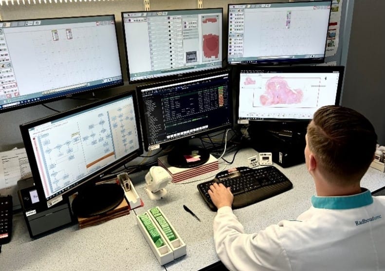
One of the big revolutions in pathology has come with the integration of digital solutions. Whole-Slide Imaging (WSI) involves scanning glass slides to produce their digital copies [4]. The first commercial WSI slide scanner was developed by James Baccus in 1994 [5]. It cost $300,000 and took approximately 24 hours to scan a single slide. Nowadays, scanning a 10×10 mm2 area in 20X takes, on average, 3.6 minutes, depending on the scanner [6].
Digital pathology has been a significant driver in the development of the discipline. It has drastically changed the field of pathology through the introduction of digital workflows, which enable highly efficient teleconsultations and digital diagnosis [7]. This transformation has also paved the way for the integration of artificial intelligence (AI) solutions . Additionally, WSI has allowed specialists to easily store archives of previous slides that can be linked to a patient’s history, thereby improving the record-keeping of previous conditions and serving as a basis for training and educating new experts.
Nowadays, pathologists leverage the knowledge gained throughout the centuries, together with innovative technologies such as WSI and AI, to bring the fields of diagnosis, prognosis, and treatment optimization to whole new levels. With the integration of novel deep learning solutions, pathologists can be supported in their assessments, resulting in faster, cheaper, and more precise results [8].
Artificial intelligence algorithms build on the foundations that WSI laid down. Deep learning solutions can automatically assess, review, and annotate the WSI slides, improving workflow and turnaround times [9]. The big breakthrough came in 2019 with the first European IVD approval of AI-based software for the detection of prostate cancer [10], shortly followed by FDA approval of the same technology [11]. Since then, AI has improved even further, and it can provide services such as automated quality control, segmentation of relevant tissues, or quantification of abnormalities.
AI-aided pathological evaluations do not have to rely on the observer’s experience, subjective interpretation, or crude semi-quantitative assessments but can rather provide reproducible and precise measurements [8]. This semi-automation allows for easier large-scale data processing in both clinical and research settings [13]. Not only does this technology bring benefits to patients, but it can also enable exponential growth in new discoveries in pathology. With increased speed and precision, researchers shift their focus from manual and monotonic assessments to the analysis of challenging cases or novel tissue structures and causes lying behind them.
By embracing these technological advances, pathology is transforming from a discipline rooted in observation to one characterized by precision and predictive possibilities. As technology continues to advance, the future holds even greater potential for enhancing our understanding of disease pathology and improving patients’ outcomes.
As a part of this revolution, Aiosyn has introduced various solutions aimed to improve diagnoses and faster discoveries. We offer products such as AiosynQC to make sure only high-quality slides are being reviewed, Aiosyn Mitosis Breast, which increases the reproducibility and efficiency of mitotic figure assessments, and our kidney image analysis services through the NephroPath platform, which offers kidney structure segmentation, quantification, and treatment response grouping, among other things. With these innovative applications and services, Aiosyn is committed to pushing the boundaries of what is possible in pathology.
Sources:


26 May, 2025 • By Diana Rosentul
19 December, 2024 • By Anna Correas Grifoll

12 December, 2024 • By Anna Correas Grifoll

25 September, 2024 • By Anna Correas Grifoll

03 July, 2024 • By Anna Correas Grifoll

18 June, 2024 • By Victoria Grosu

11 April, 2024 • By Victoria Grosu

25 January, 2024 • By Diana Rosentul
Toernooiveld 300
6525 EC Nijmegen
The Netherlands
[email protected]