AiosynQC: Automated Quality Control
We provide an AI-powered solution, AiosynQC, for automated quality control (QC) of digital histology slides in the pathology workflow.
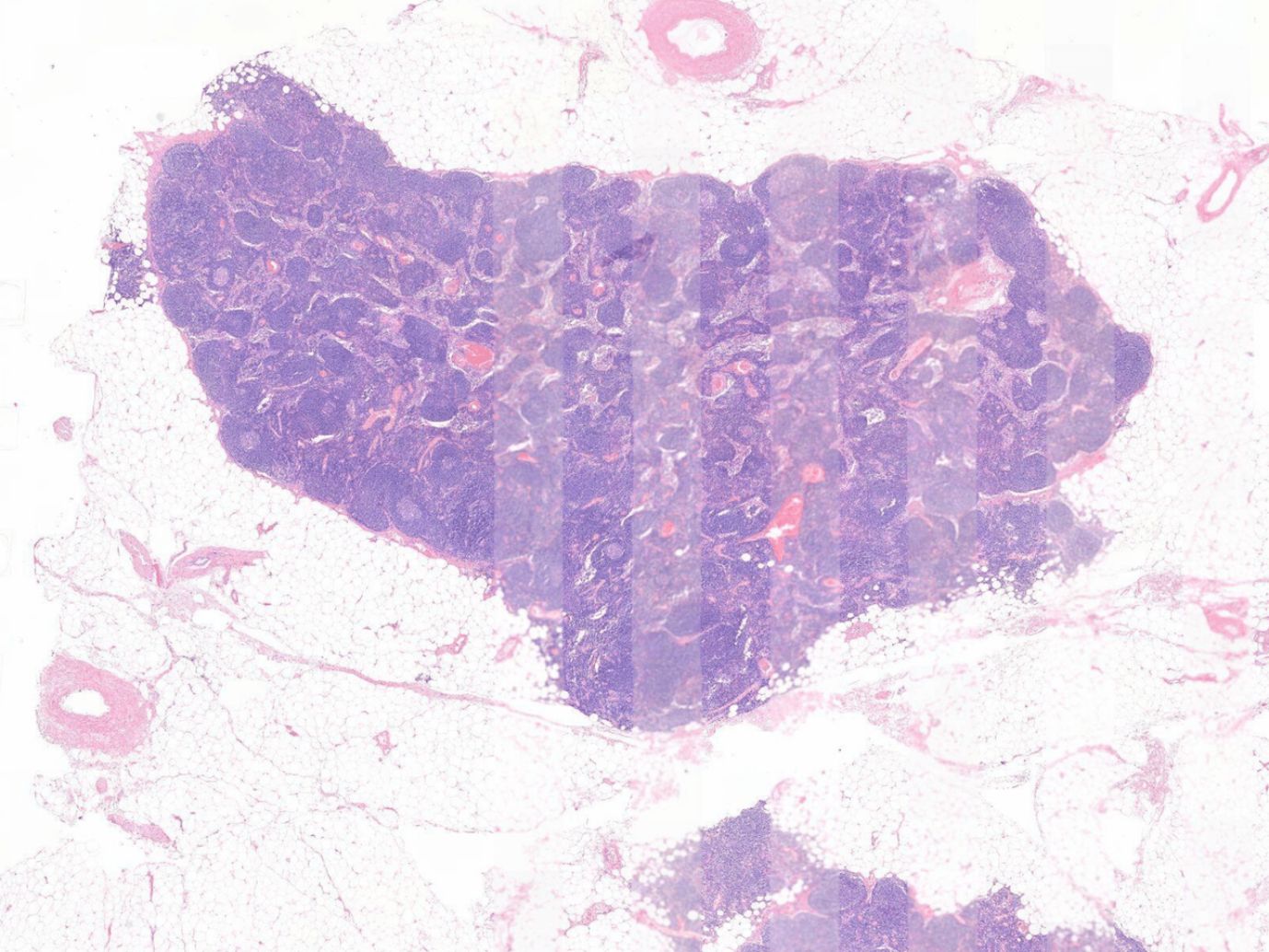
Whole-slide images (WSIs) may show reduced interpretability because of variations in pre-analytical processes and scanning artifacts. AiosynQC automates and simplifies the slide quality control process, building high-quality, efficient workflows for digital pathology laboratories and AI and data providers, ultimately supporting effective and timely diagnostics.
Improve the quality of clinical diagnostics
Improve the quality of clinical diagnostics
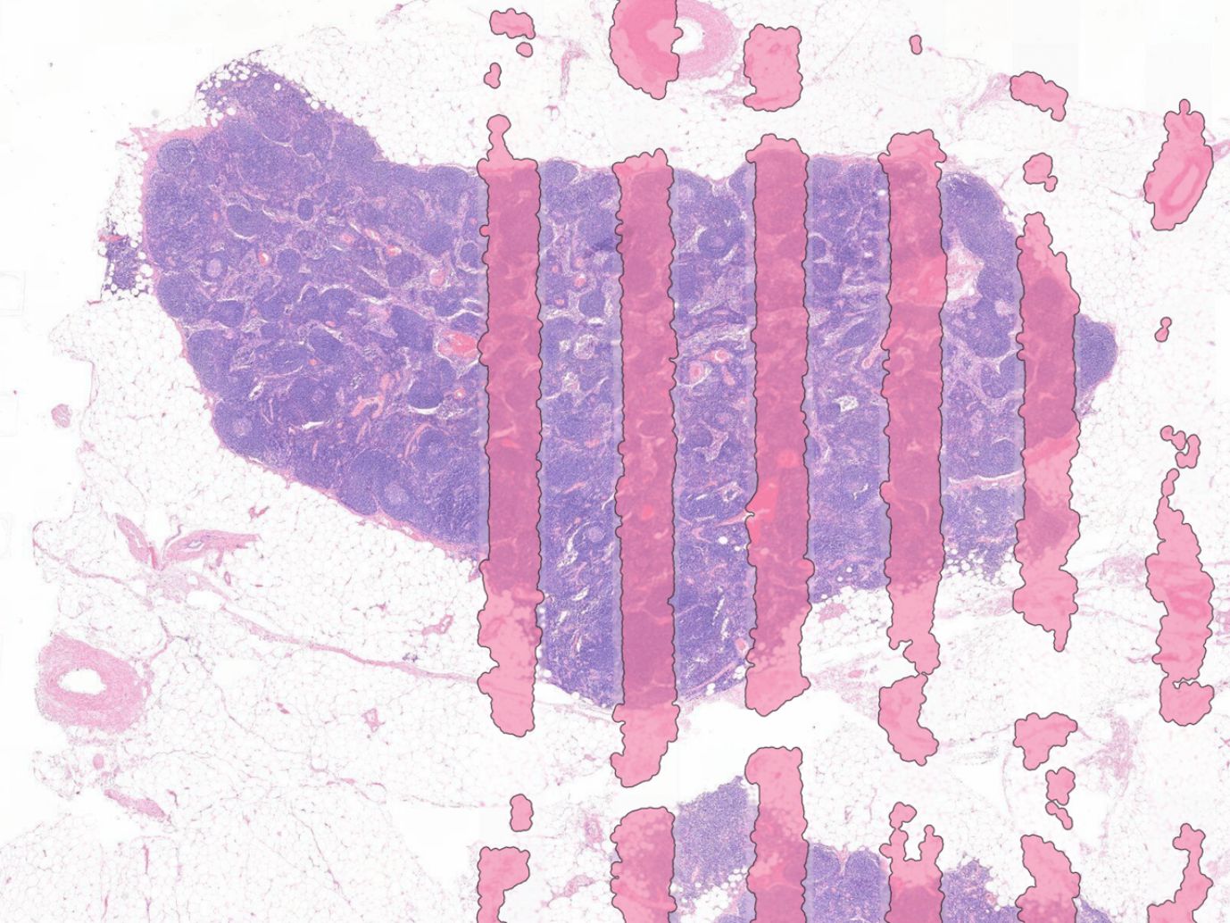
AiosynQC helps labs ensure that only high-quality images are used by pathologists. The algorithm detects and highlights the most common artifacts in H&E and IHC slides.
Reduce time spent on Quality Control
Reduce time spent on Quality Control
Automating quality control increases the efficiency of the diagnostic and research workflow by reducing the amount of time spent on manual inspection of image quality.
Start working with AI algorithms
Start working with AI algorithms
Integrating AI-powered solutions will improve your digital pathology workflow. Automating QC will ease the use of other algorithms that underperform when artifacts are present.
Integrated in your workflow
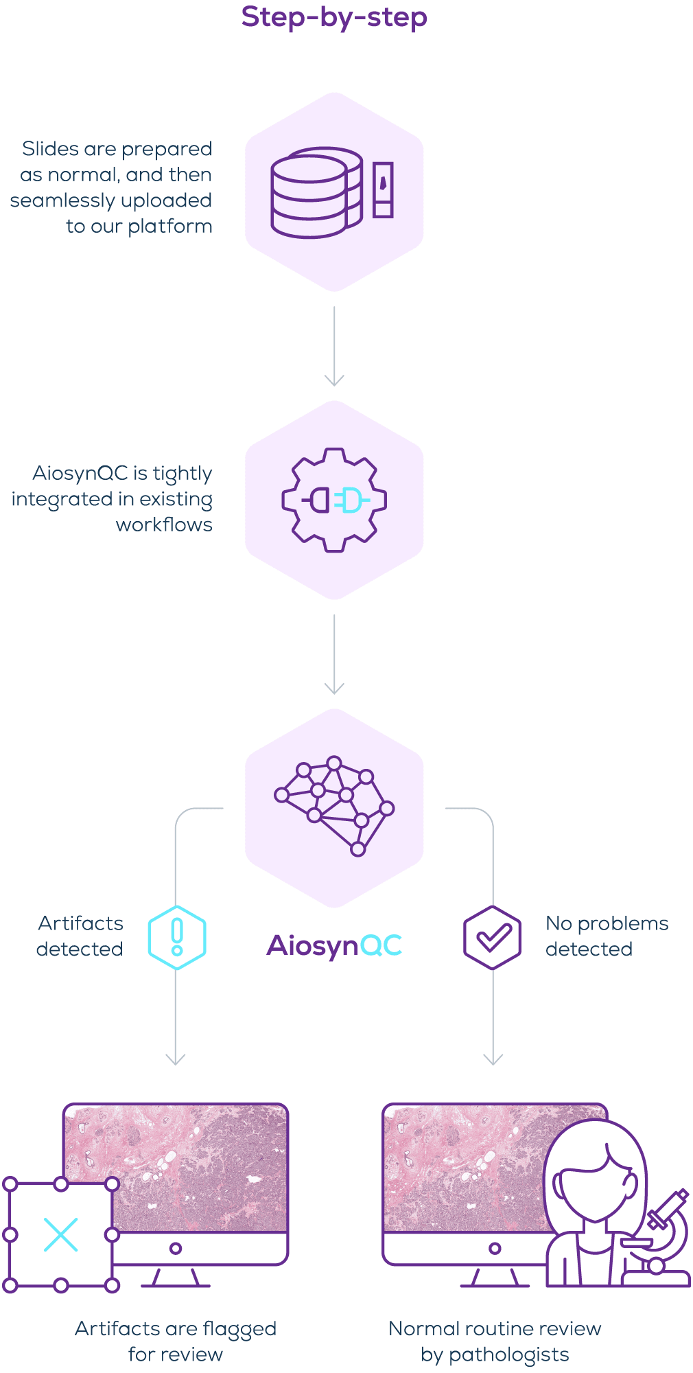
Our automated slide quality control solution integrates with major workflow providers and is available through marketplaces like Sectra Amplifier Marketplace and Paige AppLab. Slides are prepared as usual, and the algorithm analyzes images once uploaded to the platform. AiosynQC offers tailored sensitivity and customizable reporting options, adapting to the unique needs of every laboratory.
Download Product Brief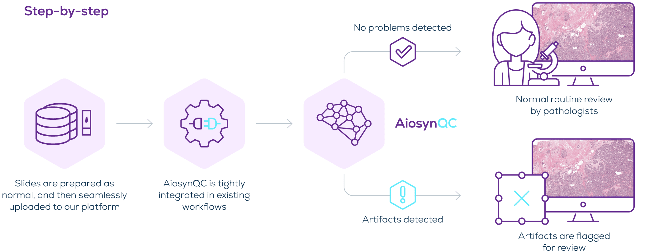
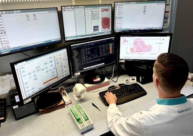
“I am excited to see AiosynQC fully integrated and seamlessly supporting our lab technicians. It is a great help to catch artifact-affected slides before they reach our pathologists without requiring zoom-ins to manually inspect the images. ”Prof. Katrien Grünberg, MD, PhD, Head of the Department of Pathology at Radboudumc
Note: In the EU and the UK, AiosynQC is not considered a medical device under European IVDR and UK MDR 2002 legislation, respectively. AiosynQC is not intended to be used as an accessory to, nor is it necessary to be used in combination with any AI or other medical devices to specifically enable them to meet their intended purpose or directly assist in their functionality. AiosynQC is for Research Use Only (RUO) in the United States or any other jurisdiction.
Discover Aiosyn’s solution for image Quality Control
AiosynQC automatically recognizes common artifacts and classifies slides based on your lab’s acceptance criteria. Tightly integrated into your existing workflow, this application advances clinical diagnostics, prevents delays, and makes the digitization of large cohorts much easier.
Schedule a demo to discover how AiosynQC works on your images and how it will improve your digital pathology workflow.
Book a demoAiosynQC news and articles
-

Aiosyn expands AI-powered quality control with incomplete scan detection
09 April, 2025 • By Anna Correas GrifollRead more -

Techcyte and Aiosyn collaborate to integrate AI-powered slide QC and mitotic counting into the Fusion™ digital pathology platform
20 March, 2025 • By Anna Correas GrifollRead more -

Case Study: Automating Radboudumc’s digital slide Quality Control with AiosynQC
19 December, 2024 • By Anna Correas GrifollRead more
Questions? We are happy to answer
"*" indicates required fields
The most frequent questions about AiosynQC
Is AiosynQC limited to hematoxylin and eosin (H&E) staining or it can assess immunohistochemistry (IHC) slides as well?
AiosynQC supports both H&E and IHC staining.
Does the tool only classify a slide image as having or not having artifacts, or does it also segment the area of the artifact?
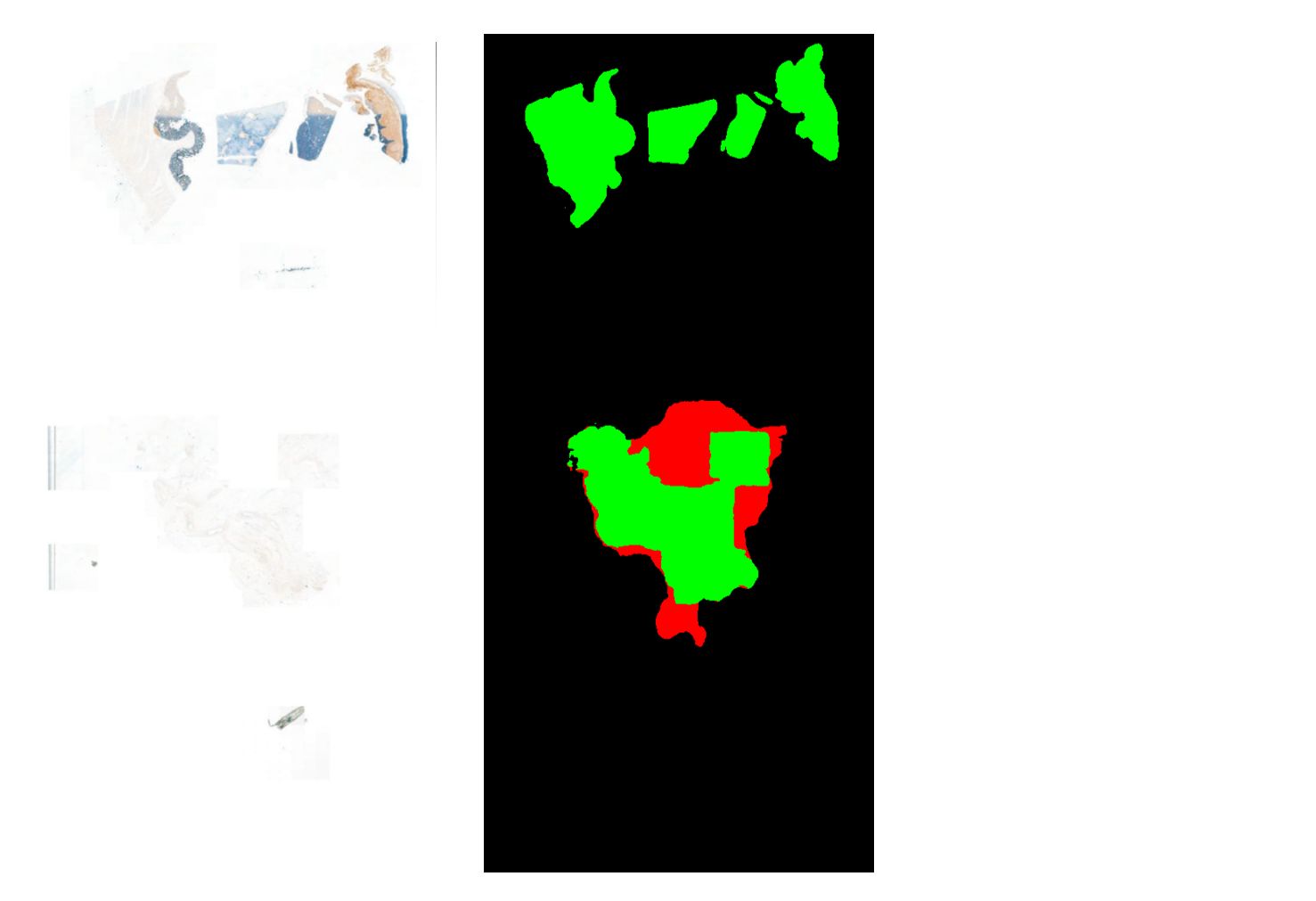
AiosynQC detects the most common quality artifacts and segments the affected areas.
What types of artifacts can the software detect?
AiosynQC can detect the most common quality artifacts in whole slide images (WSIs), including out-of-focus areas, incomplete scanning, air bubbles, tissue folds, pen markers, dust, white balance problems, and ink.
How does the output of your models look, and how can it be customized to specific workflows?
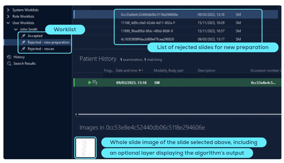
AiosynQC can provide results in multiple formats, including worklists of accepted and rejected slides, detailed quality scores, and color-coded indicators that highlight the presence or absence of artifacts. For a more in-depth analysis, users can click on a whole slide image to view an overlay that segments affected regions and indicates the artifact types. The output and reporting of compromised slides can be tailored to meet the specific needs of the laboratory. For example, labs can configure the system to flag only tissue folds exceeding a certain size threshold while ignoring smaller ones.
What file types are compatible with AiosynQC?
The tool is compatible with OpenSlide and supports various standard formats such as TIFF, SVS, MRXS, and more.
Can AiosynQC be run on-premise?
Yes, the software can be run on-premise as well as in the cloud.
Is the software CE-marked?
AiosynQC is not CE-marked because, under the European IVDR, it is not considered a medical device in the EU, nor is it intended to be used as an accessory to any AI or other medical devices.
What percentage of slides are flagged by AiosynQC?
The incidence of artifacts varies depending on the laboratory workflow and equipment. AiosynQC offers tailored sensitivity and customized reporting options to adapt to the specific needs of each laboratory. For instance, labs may choose to flag images with significant anomalies while ignoring small artifacts that typically do not compromise readability. However, other centers may prefer a more comprehensive review to ensure all images sent to the pathologist are of high quality and do not require a rescan.