
Custom AI pathologyfor your study, lab or product
Whether you have a new research question, an experimental stain, or a novel endpoint, our AI experts can help you build a tailor-made solution. Custom AI pathology software developed on your own data and delivered per study or ready for production use.
- Developed on your data
- Validated on your endpoints
- Delivered per study, or for production use
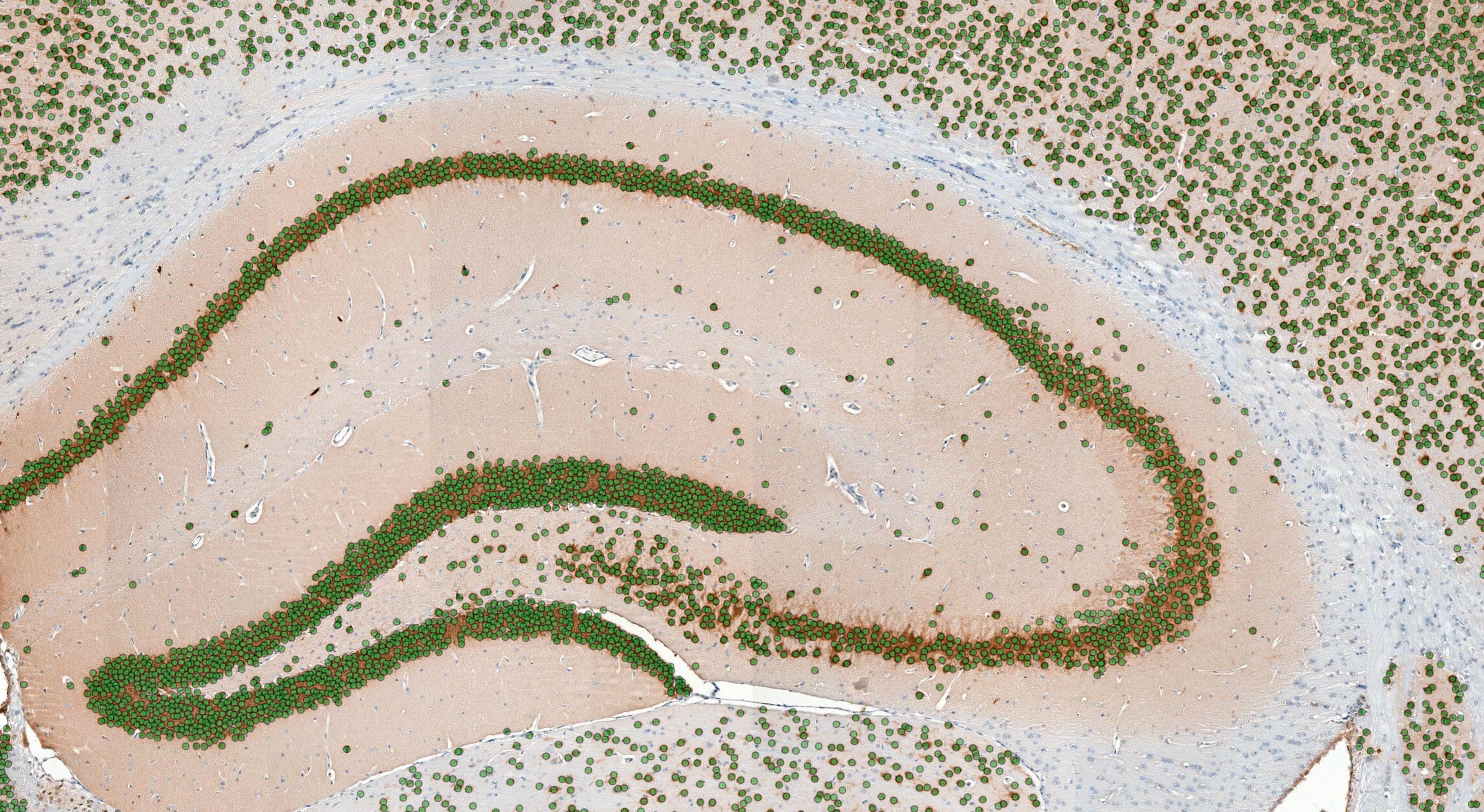
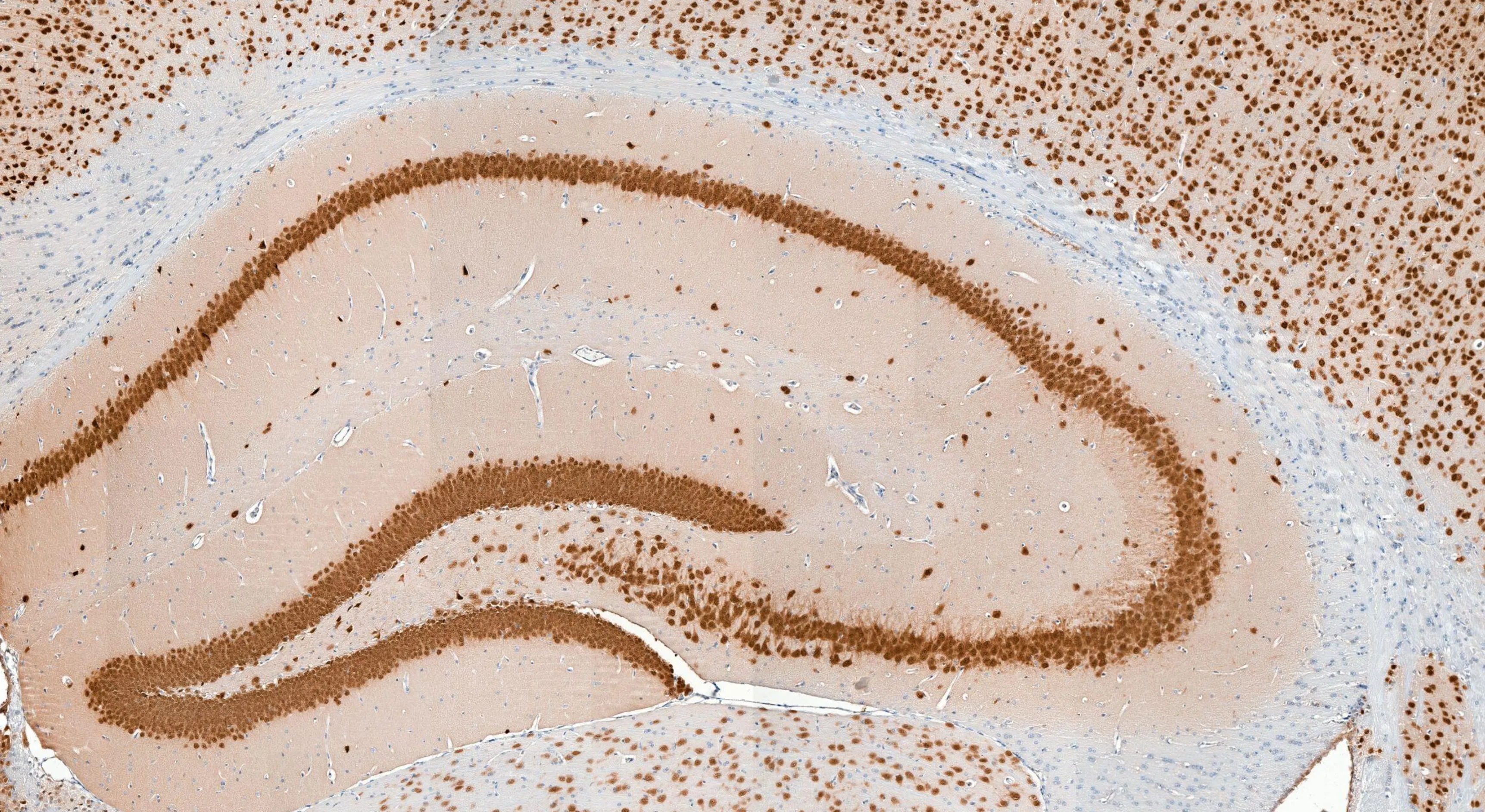 Brain sectionDetected neurons
Where innovation meets expertise
From idea to production, each customer journey is unique. However, we have developed a method that streamlines this journey, enabling custom AI pathology development as a service.
Use cases
For a study
Conduct a study on a new endpoint, tissue, stain or species. Defined by your own protocol, the model is built on your data and read-outs come back as a quantified report.
Biopharma read-outsFor a laboratory
AI-powered automation and quantification that your laboratory needs in routine use, integrated in the viewer and image management system you already have.
Clinical solutionsFor a product
A vendor-neutral, production-ready, AI-powered solution behind your pathology product. Built as a reliable component that is deeply integrated within your infrastructure.
Discuss an integrationInnovation as a service
The Aiosyn team has decades of experience building AI-powered software solutions for digital pathology, for both academia and industry. Let us help you.
Stage 1 · Scope
Tell us what you need
- Endpoints, tissue, stain and species to be analyzed
- The reference you want to compare against
- Feasibility & benchmark against the models we already have
- Support in building the business case
Stage 2 · Develop
Build and validate your solution
- Customized model development for your own data and endpoints
- Annotation service with expert pathologists
- Models trained and validated on unseen data: avoid domain shift
- Reviewed in our online viewer, alongside your own images
- Offload technical work to our AI experts, and focus on your science
Stage 3 · Production
Flawless operation within your workflow
- Technical specifications and an implementation plan with your team
- Automatic data transfer, read-outs in your own viewer or product
- A gradual increase toward full workload
- On-premise within your network, or in the cloud
- Monitoring and continuous improvement on your feedback
Validated before it is relied on
Every analysis we run was developed together with expert pathologists and validated on real cohorts before it was used on a study. A custom model follows the same route.
Method validation
Across 80 cases, AI lesion-area read-outs matched expert pathologist scoring
Developed with TNO for a read-out the suite did not cover: atherosclerotic lesion area across the full aortic-root cross-section of every animal in the cohort.
Atherosclerosis · aortic-root cross-sections
Explore atherosclerosis read-outsThree case studies, each one customized
Each solution is unique and customized, from research idea to publication or production.
For a study
Kidney damage quantified in a diabetic model
Physiogenex assessed dapagliflozin efficacy in a diabetic nephropathy cohort with NephroPath. The automated read-outs resolved a treatment effect that classical semi-quantitative scoring did not.
Read the case study
For a laboratory
Quality control on 1,200 slides a day
Radboudumc digitizes around 1,200 slides daily, and inspecting them for artifacts by hand would add more than ten hours of work a day. AiosynQC checks each one as it arrives.
Read the case study
For a product
Kidney AI running inside another vendor’s scanner
Aiosyn’s kidney models run at the edge inside Pramana’s scanners, detecting structures inline as the slide is digitized (real time) rather than in a batch afterwards.
Read the announcement
Frequently asked questions
How is our data handled?
You can upload whole-slide images to our secure platform, where they are accessible to model development. Other transfer methods can be accommodated, discussed during scoping.
How many cases do you need?
Each project has unique challenges and requirements, and needs careful examination. However, a rule of thumb is that we can usually build a proof of concept (demonstrator or minimum viable product) with 50-100 whole-slide images, sometimes even less.
How long does it take?
If an existing model applies to your images, read-outs can be ready within a week. For a fully custom project, timelines are discussed during the scoping phase and can range from weeks to months.
Tell us what needs measuring
Describe the tissue, the stain and the endpoint, or the read-out your own product is missing. We will tell you whether an existing model covers it, or we will build it for you.