
Atherosclerotic lesion analysisacross the aortic root
Aiosyn's AI-powered image analysis quantifies atherosclerotic lesion area across the full aortic-root cross-section of every animal in your cohort. Every read-out is returned as consistent, quantitative measurements, supporting the statistical power of preclinical studies.
- Fully measure every section
- Consistent measurements across runs
- Validated against expert pathologist scoring
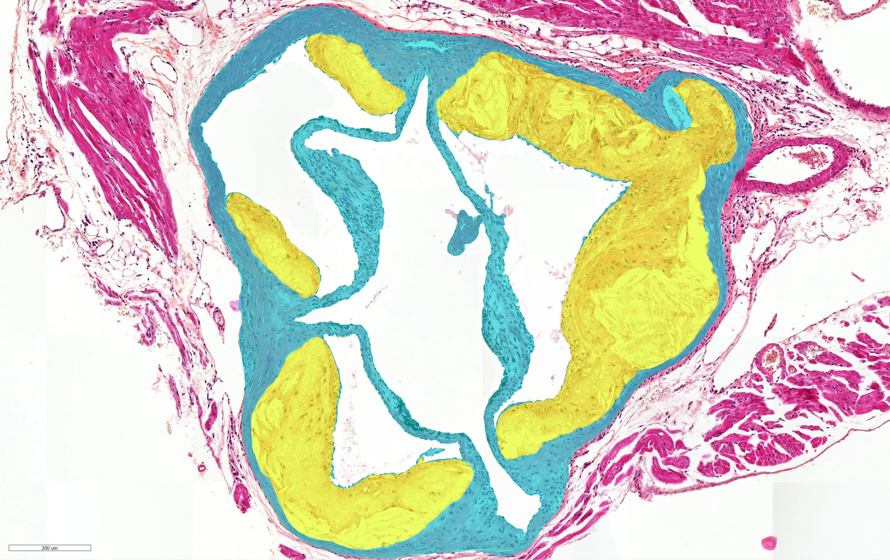
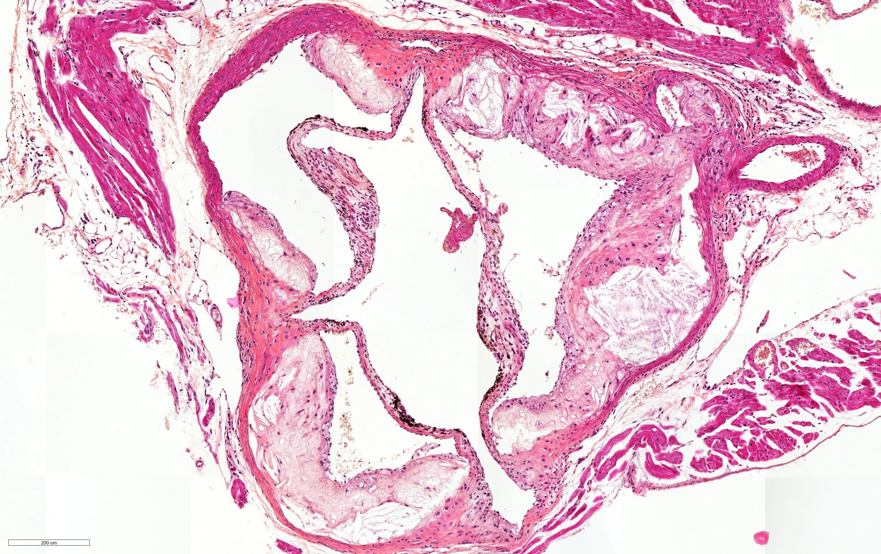 Stained sectionAI read-out
- Atherosclerotic lesion
- Vessel wall
Agreement with expert pathologist scoring
A method validation with TNO, not a published paper: an expert pathologist measured the lesion area on each mouse aortic-root cross-section, and the AI measured the same sections independently. The kidney work is the published one.
AI against expert pathologist
Lesion area, mouse aortic-root cross-section
- Correlation
- r = 0.96
- Variance explained
- R² = 0.91
- Sections
- 80
Each point is one cross-section: the pathologist's lesion area on the horizontal axis, the AI's on the vertical, against the line of perfect agreement.
What comes back from every section
An aortic root is normally read by eye, from a few sections, onto a grading scale. Aiosyn delineates the atherosclerotic lesion and the vessel wall on every section in the cohort, and measures the lesion area from those delineations.
- Lesion area per cross-section, in µm², measured across the full section rather than a sampled part of it.
- The vessel wall delineated on the same section, so the lesion is measured against the wall it sits in.
- The delineations themselves, so every figure traces back to the tissue it came from.
On mouse aortic roots
Our measurements are validated on mouse aortic-root cross-sections. For analysis on different vessels or species, check out our custom AI development.
- 1
Share your data
Whole-slide images of aortic root sections in your preferred format.
- 2
Measure
Automated analysis, allowing for a quick turnaround.
- 3
Report
A comprehensive report of our analysis is shared, including the raw data for further study.
Plaque composition
Lesion area answers how much. Composition answers what the lesion is made of.
We quantify composition on your sections as part of a scoped engagement, alongside the lesion area the standard suite returns.
Smooth muscle cells and macrophages are measured in MAC3, the necrotic core in HPS, and fibrous area in Picrosirius Red.
If composition is the endpoint your study turns on, tell us which components you need measured and we will tell you where the model stands on them.
Co-developed with TNO Health & Work
Through our partnership, we co-develop these models together with TNO. Their preclinical and pharmaceutical research expertise sits behind the study design and the tissue, our AI behind the read-outs, and the models are applied within TNO's preclinical studies with more study areas in development.
Continue with another tissue
Biopharma overview- AtherosclerosisLesion area, whole cross-section
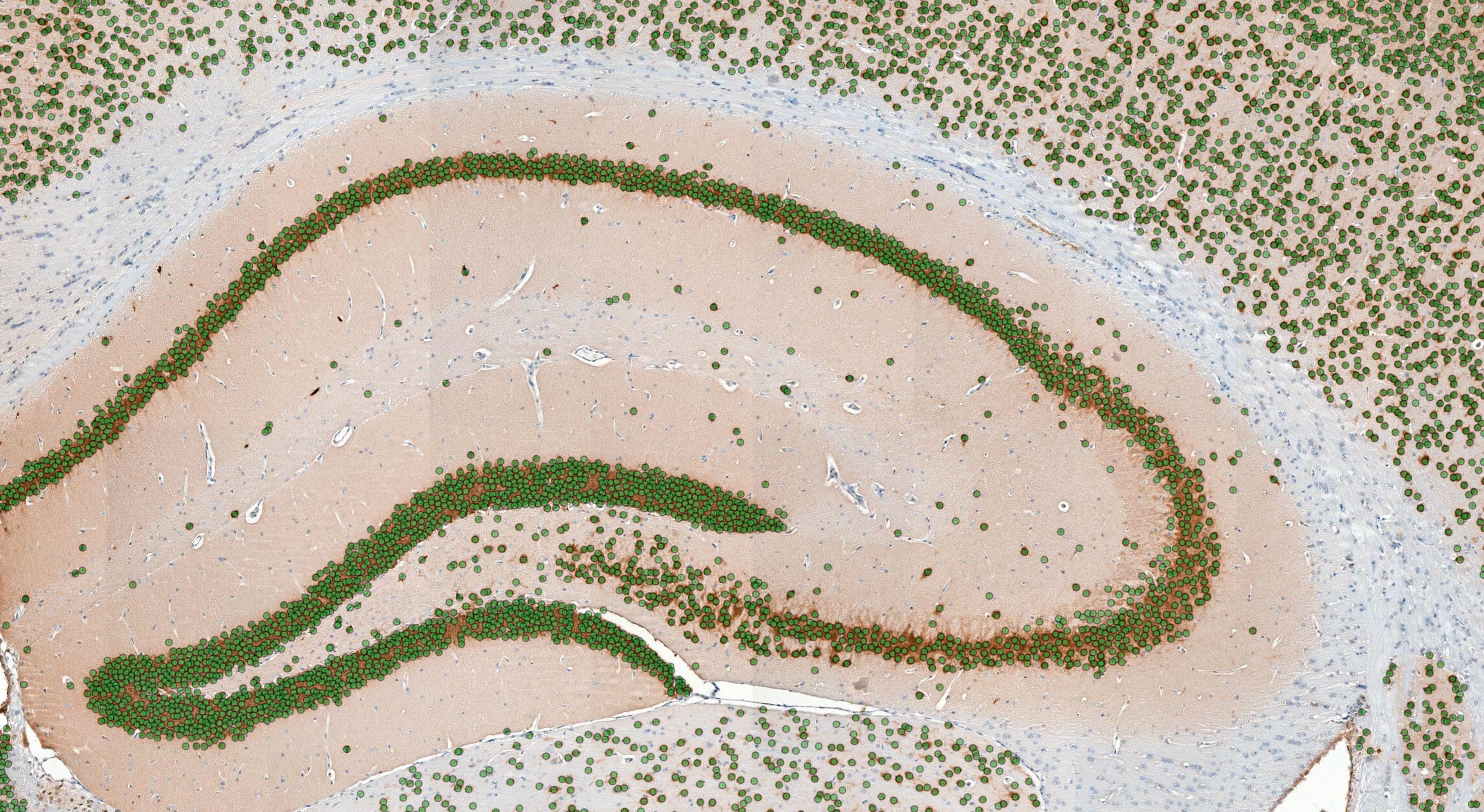 BrainNeuron density per region
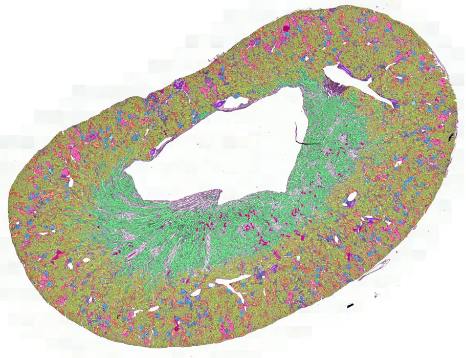 NephroPath (kidney)Every glomerulus, scored
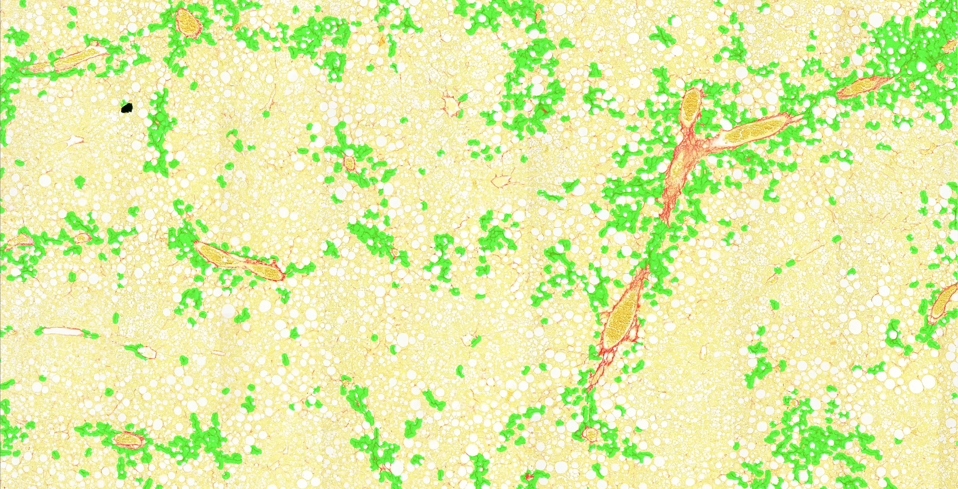 LiverFibrosis extent
Put it on your own aortic roots
Send a handful of aortic-root sections and we will return the lesion areas on your own tissue, so you can judge the measurement before a study depends on it.
Message sent
Thank you. We will get back to you.