Aiosyn Mitosis Breast: Mitotic figure counting
We provide an IVDR-certified mitosis AI application to aid clinical laboratories with the identification and counting of mitotic figures in breast H&E sections.
The manual process is time-consuming and subject to variability, and there is a need for solutions to increase its efficiency and consistency of results. Aiosyn Mitosis Breast [1] uses deep learning to detect mitosis in breast whole slide images, saving pathologists time and providing laboratories with a more standard process for assessing tumor growth.
1. Aiosyn Mitosis Breast, for use in clinical diagnostics, has obtained CE-mark certification under the EU IVDR. In addition to Aiosyn Mitosis Breast, we offer Aiosyn Mitosis Research for biopharma and research institutions.
Up to 60% time savings
Up to 60% time savings
The algorithm helps laboratories improve the process of mitosis analysis. In a clinical performance study, pathologists achieved up to 60% time savings per slide with Aiosyn Mitosis Breast.
32.6% enhancement in consistency
32.6% enhancement in consistency
Aiosyn Mitosis Breast has been shown to reduce variability among observers, leading to a 32.6% improvement in consistency between pathologists.
AI integrated in your workflow
AI integrated in your workflow
Aiosyn Mitosis Research and Aiosyn Mitosis Breast are offered as a modular software solution that can be integrated into existing workflows, removing the need to adopt a different viewer.
Your workflow with Aiosyn Mitosis Breast
Our AI-powered solutions integrate with major workflow providers and are available through marketplaces like Sectra Amplifier Marketplace and Paige AppLab. Slides are prepared as usual, and the algorithm analyzes images once uploaded to the platform.
Over 90% of surveyed pathologists who tried Aiosyn Mitosis Breast would like to use the algorithm in their daily practice.


Improving mitosis counting for clinical diagnostics and biopharma
-
Aiosyn Mitosis Breast for clinical diagnostics
-
Aiosyn Mitosis Research for biopharma
-
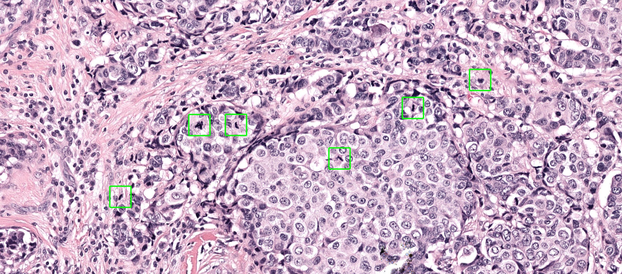
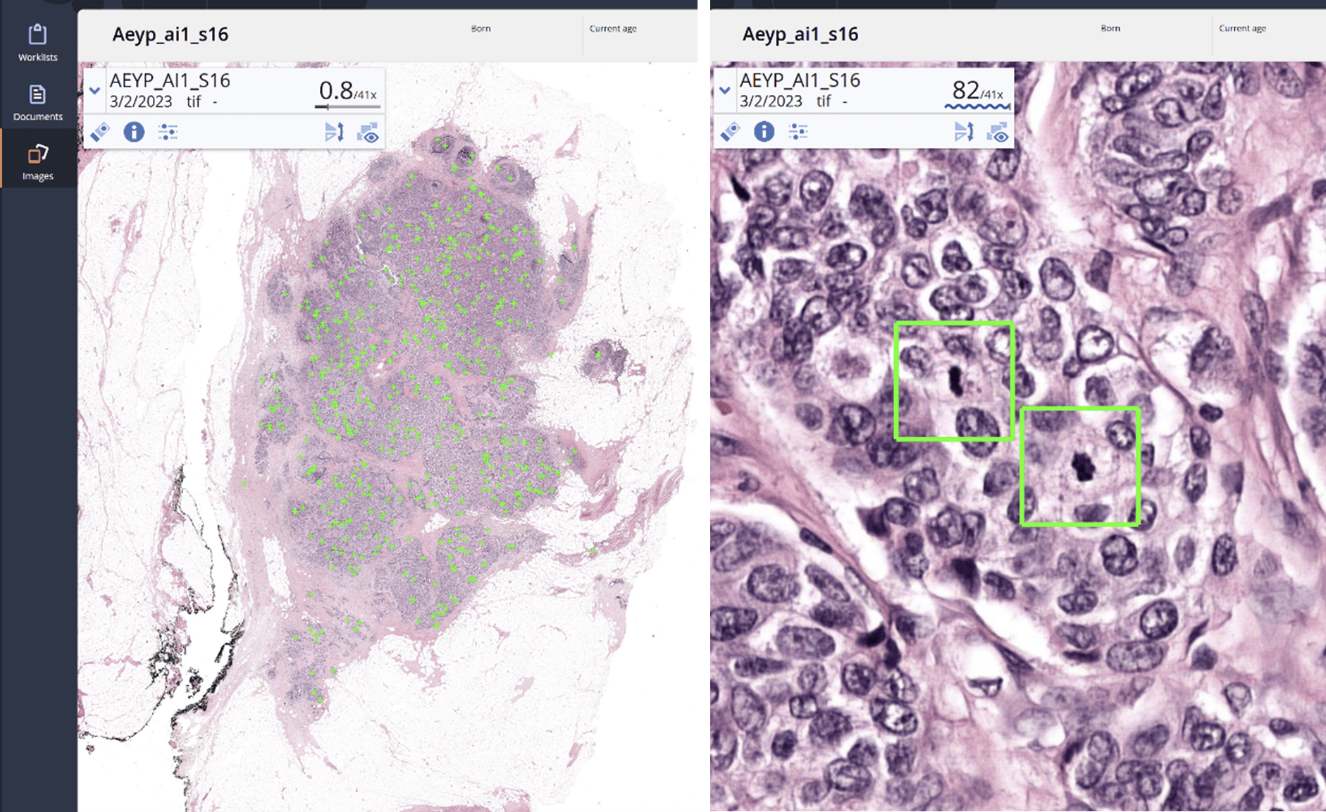
Aiosyn Mitosis Breast assists pathologists in detecting mitotic figures in whole slide images of breast biopsies and resections. Mitoses are detected and highlighted by the algorithm before the pathologist opens the WSI, thereby indicating areas with mitotic activity and assisting in the quantification.
In a clinical performance study involving 28 pathologists from 9 countries, users achieved up to 60% time savings per slide when supported by Aiosyn Mitosis Breast. On average, pathologists showed a 15.5% increase in productivity when reviewing resections. Additionally, the algorithm lead to a 32.6% improvement in consistency between pathologists.

-
Aiosyn Mitosis Research aids research labs with the identification of mitotic figures in H&E sections. With improved and reproducible detection, new biomarkers can be discovered and researched more accurately.
Automated whole-slide level assessment enables the processing of caseloads that would not be possible with human annotators. By streamlining the process, Aiosyn Mitosis Research can assist laboratories in adopting a more consistent and efficient protocol for the identification of novel pathology-based biomarkers, resulting in improved and standardized results.

Aiosyn Mitosis news and articles
-

Aiosyn and Telemis advance breast cancer diagnosis with IVDR-certified AI technology
26 June, 2025 • By Anna Correas GrifollRead more -

Aiosyn Mitosis Breast becomes the first AI-powered mitosis detection solution to achieve CE mark certification under IVDR
07 January, 2025 • By Anna Correas GrifollRead more -

AI-assisted mitosis counting in breast cancer. A large-scale validation study.
25 March, 2024 • By Jeroen van der LaakRead more
Questions? We are happy to answer
We will get back to you as soon as possible.
"*" indicates required fields
The most frequent questions about Aiosyn Mitosis Breast
Is the algorithm CE marked?
Yes. Aiosyn Mitosis Breast has obtained CE-mark certification under the EU IVDR.
How does it compare to standard counting?
The algorithm automatically detects mitotic figures before the case is reviewed. When the WSI is opened, all identified mitoses are highlighted, helping pathologists quickly locate high mitotic activity areas and assist in the count. With Aiosyn Mitosis Breast, pathologists can review the algorithm’s output instead of manually identifying mitoses, reducing evaluation time by up to 60%.
Does the tool support hotspot detection?
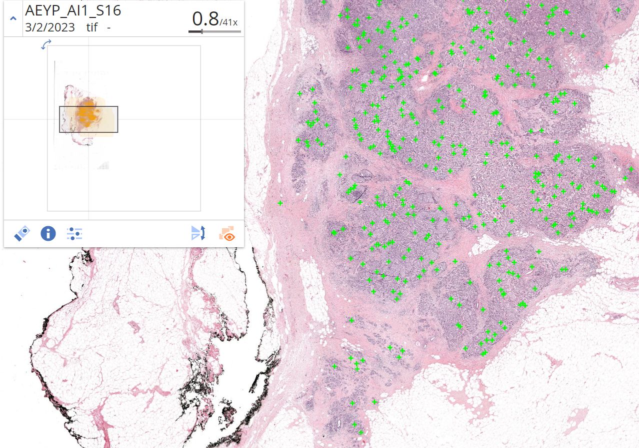
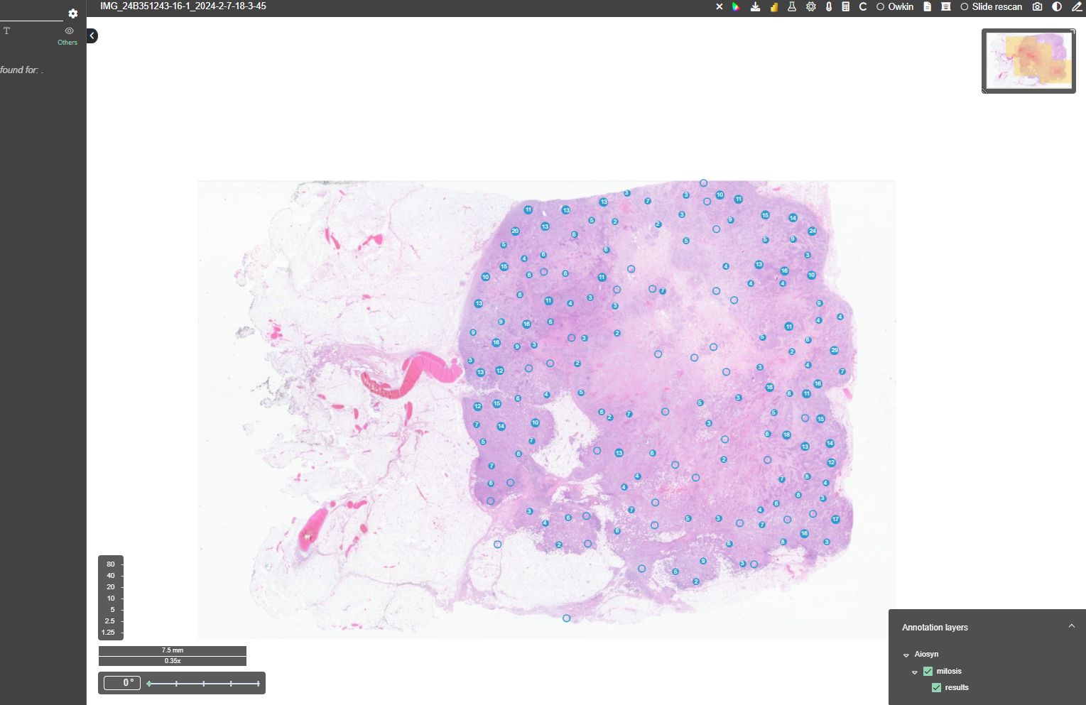
Aiosyn Mitosis Breast provides the coordinates and highlights the mitotic figures. This data allows pathology viewers to display mitotic activity, assisting pathologists in identifying areas of high mitotic density. This supports both hotspot detection and accurate counting of mitotic figures. The specific visualization features (heatmaps, squares, etc.) for these high-activity areas vary by the viewer. We are also working on integrating a hotspot detection feature into the clinical version, which is currently only available in Aiosyn Mitosis Research.
What data and information are required to conduct a trial period?
You can request a demonstration of Aiosyn Mitosis Breast very easily. First, you upload a few whole-slide images to our secure online platform. Then, we apply the solution to detect mitotic figures throughout the image. Finally, we give you access to our online viewer where you can examine the product’s detections together with your images.
What are the IT requirements for integrating Aiosyn Mitosis Breast into our existing infrastructure?
We are a flexible provider of AI-powered solutions, and can accommodate most setups, with two common deployment options as follows. First, performing the integration via an existing marketplace, for example: Sectra Amplifier Marketplace, Paige AppLabTM, PathAI AISight, Proscia, etc. We work with most manufacturers to bring our products to your lab with ease. Second, integrating directly with your IMS or custom system via simple HTTP(S) API. In this case, we require that your system is able to send us notifications when a new image is available for processing, and later displaying the detections of the product (mitotic figures). Both deployment options are available on-premise (installed in your own hardware inside your network), or in the cloud (we use tier-1 infrastructure from AWS and Azure).
How does this solution differentiate itself from other AI solutions available on the market?
Aiosyn Mitosis Breast is the first AI-powered mitosis detection solution to achieve CE mark certification under IVDR. The application went through a rigorous validation and multi-center clinical performance study which demonstrated significant improvements in both efficiency and consistency among 28 pathologists across 9 countries when using it. Over 90% of surveyed pathologists who tried Aiosyn Mitosis Breast would like to use the algorithm in their daily practice. In addition, the solution is modular and can be integrated into existing workflows, minimizing investment and disruption of current processes.
What is the scaling capability for handling large cohorts of slides?
For large volumes, Aiosyn Mitosis Breast computing resources can be deployed in the cloud. This has the benefit of providing virtually unlimited scaling capabilities. When needed, extra hardware resources can be invoked to process a large number of slides in parallel, substantially reducing turnaround time. These resources are discarded after the workload is completed, limiting processing costs.
Is the algorithm compliant with GDPR and other data privacy regulations?
Yes, Aiosyn Mitosis Breast is compliant with GDPR and other standard regulations.
What are the available pricing models? Is there an option for per-slide pricing or is it subscription-based?
We establish the pricing models based on your needs and preferences. Both subscriptions and per-slide pricing are available.