NephroPath platform
Through the NephroPath platform, we provide a detailed evaluation of histological biomarkers in preclinical and clinical kidney samples1. Our AI-based quantification offers reproducible, consistent, fast, and more detailed scoring of renal pathology biomarkers compared to traditional pathologist scoring.
Explore our image analysis services and partner with us to improve your (pre)clinical studies.
1. The NephroPath platform is for Research Use Only and should not be used for diagnostic procedures.
Download Report Sample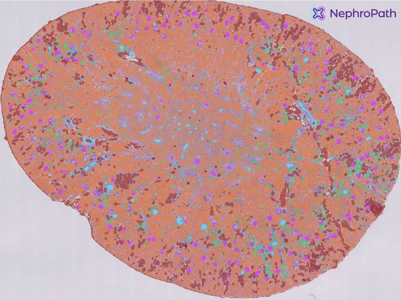

AI-assisted image analysis services
-
Standard whole kidney analysis
-
Custom kidney analysis
-
Enrich your study with AI-powered whole kidney tissue quantification. We offer insights into your data through:
- Accurate segmentation and delineation of relevant kidney structures
- Extent of glomerulosclerosis and fibrosis
- (Immune) cell quantification, including spatial relation to tissue
- Online/offline access to all results

-
Leverage Aiosyn’s expertise in AI and the latest technology to create a tailored analysis for kidney tissue. We offer development and prototyping of robust and performant models with a quick turn-around time:
- IHC/Biomarker quantification in specific animal models
- Treatment response grouping
- Tissue microenvironment analysis
How can we assist your team?
We fully automate the evaluation of renal pathology images, addressing key challenges by providing:
- Detailed reports on kidney tissue analysis swiftly upon data submission.
- Precise quantification to improve the reproducibility of results.
- Improved objectivity of pathologist’s assessments in multi-center (pre)clinical studies.
- Customized projects for new and existing biomarkers.
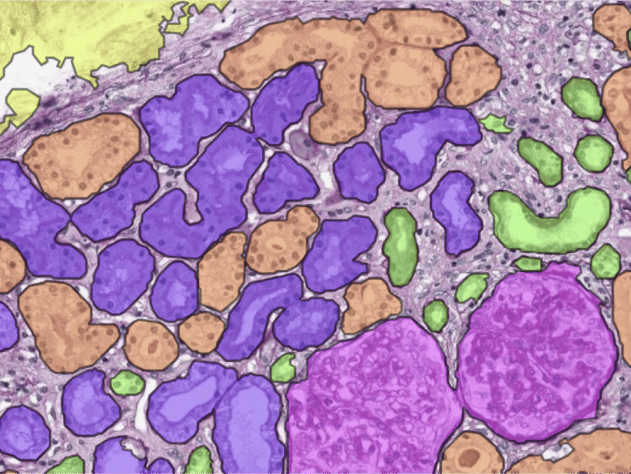
Segmentation of main tissue classes
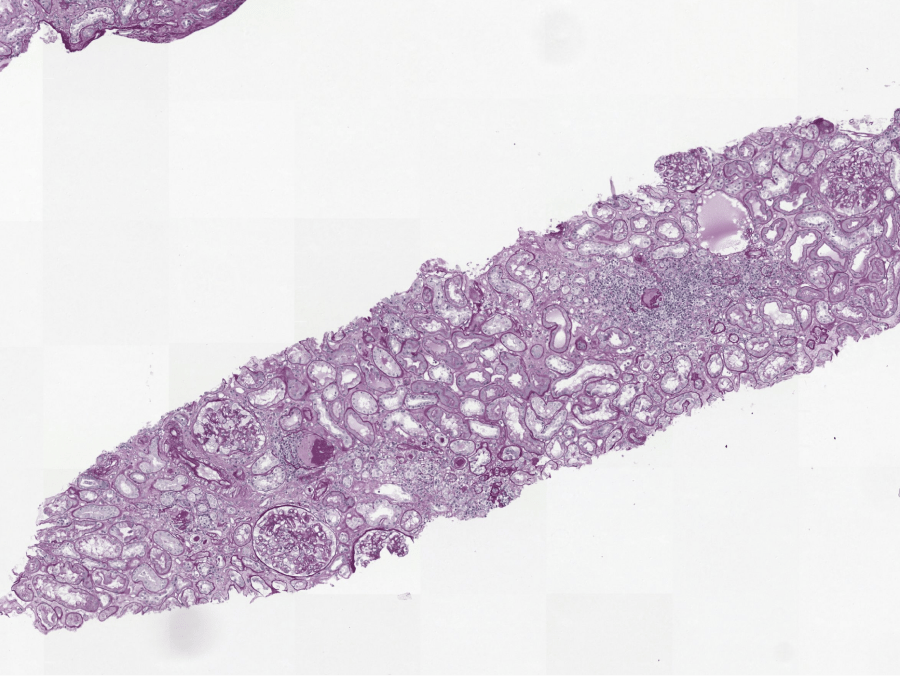
Our AI can accurately segment relevant kidney structures, among which: (sclerotic) glomeruli, proximal/distal/atrophic tubuli, arteries & capsule. Leveraging this data, we can provide a wide variety of quantitative and spatial measurements, such as the number of glomeruli in a biopsy or the tissue area consisting of interstitium.
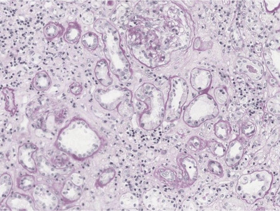
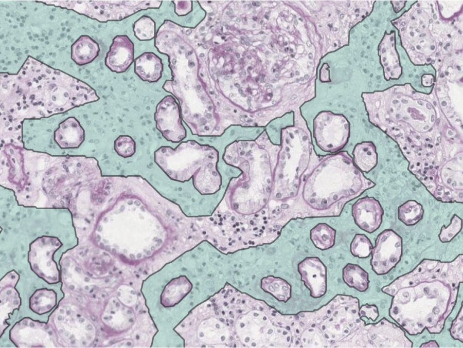
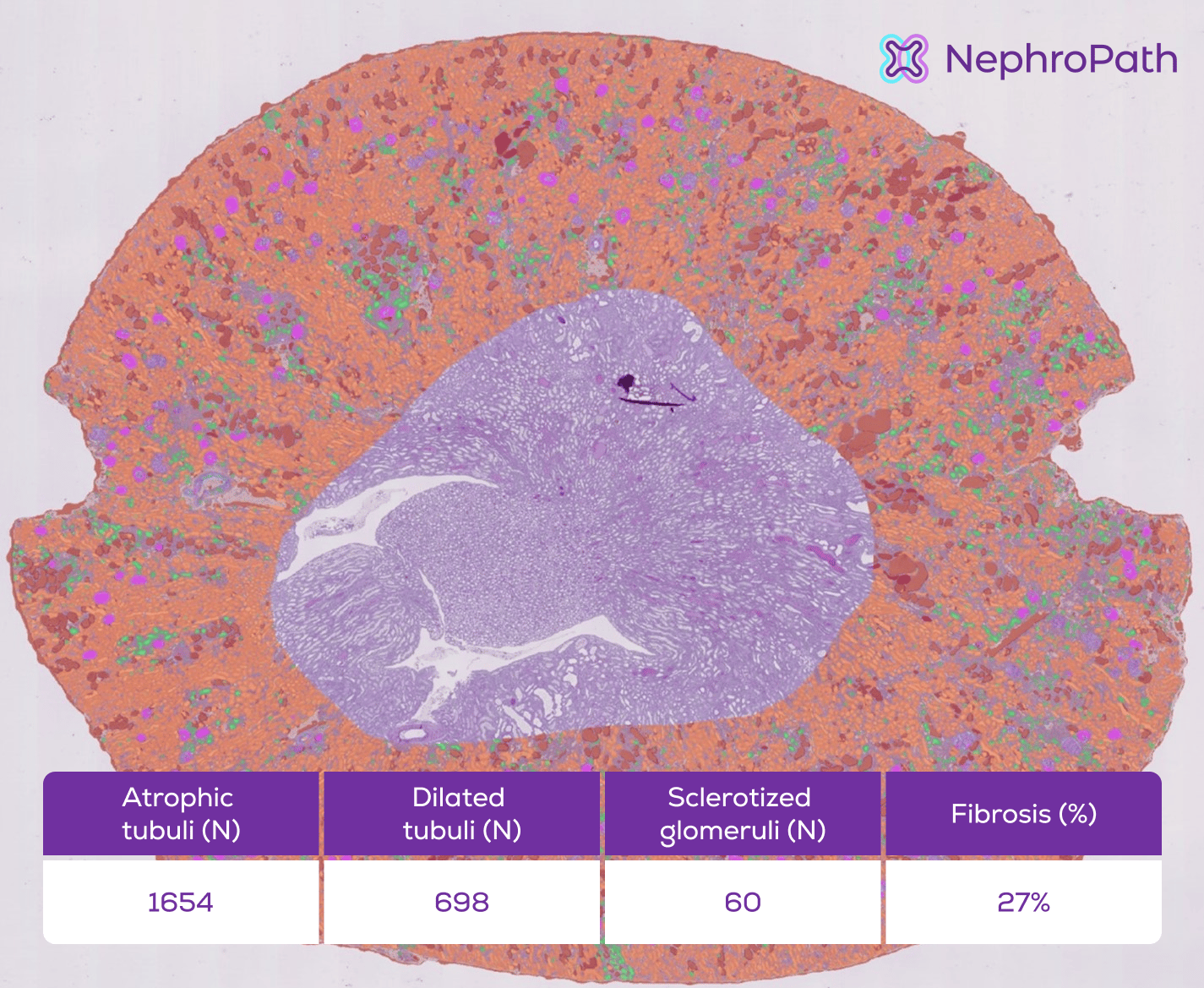
AI-Powered quantification of fibrosis
Renal fibrosis is a key biomarker for CKD progression and for prognosis of kidney transplantation. Current scoring systems are based on crude semi-quantitative assessments, limited to the capability of the human eye. AI-based fibrosis quantification yields an objective and precise measurement that can aid pathologists and biopharma services with their assessments.
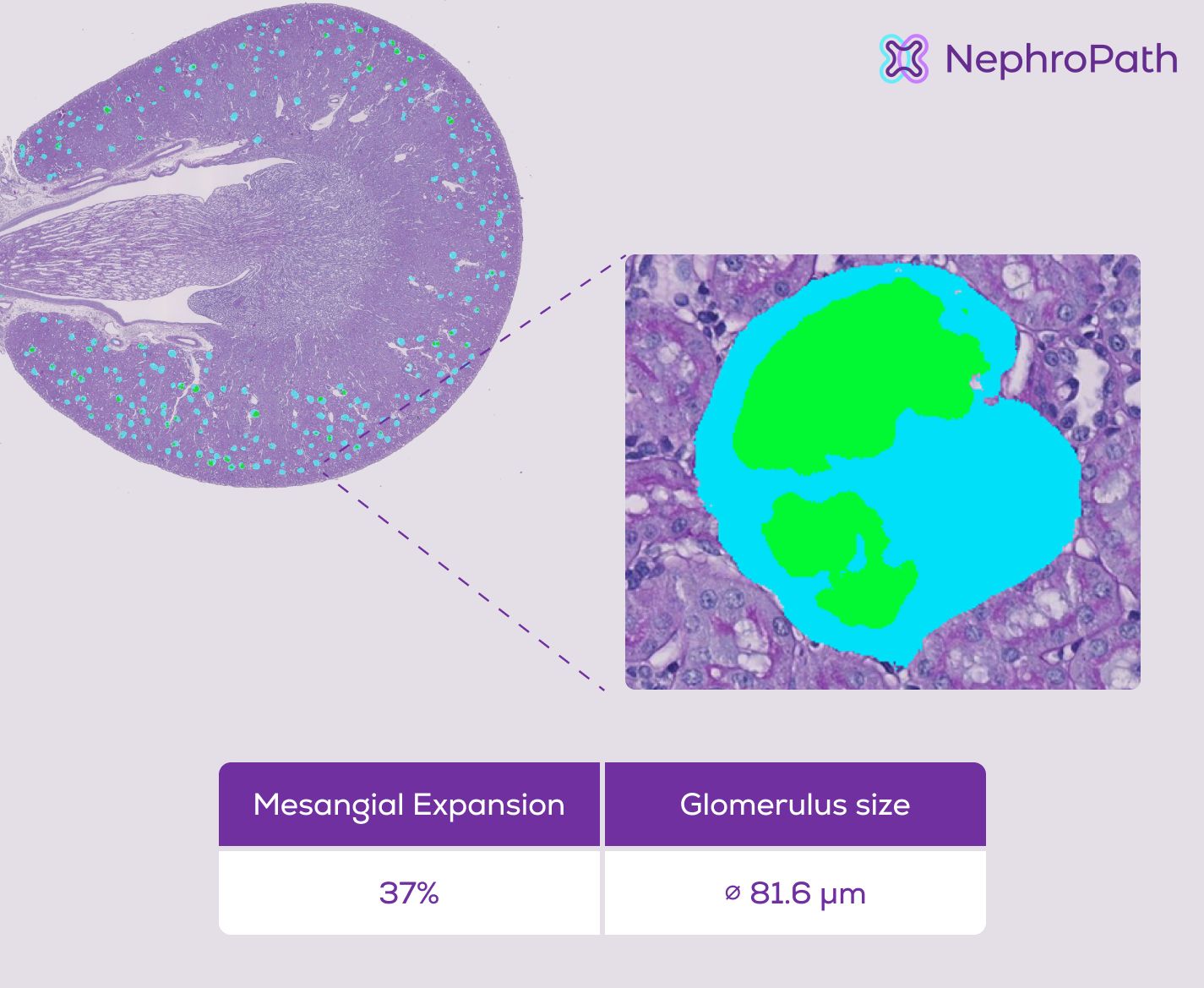
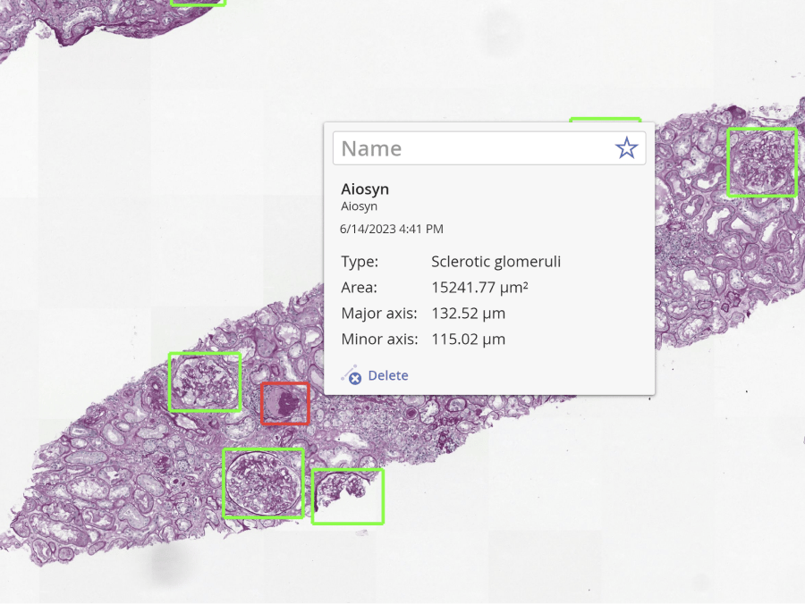
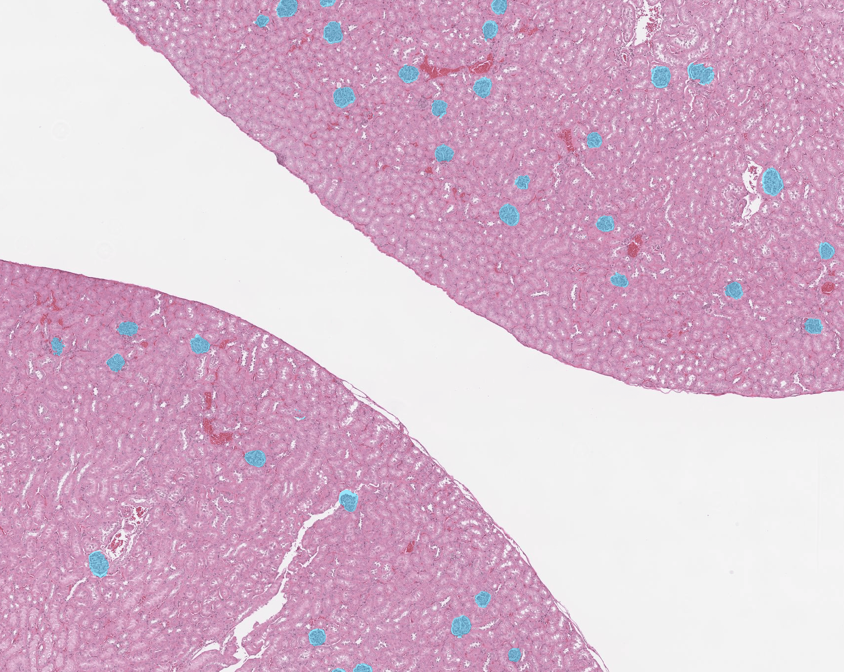
Localization and counting of glomeruli
The assessment of glomeruli is a quality assessment of every renal sample. NephroPath GlomeruCount provides a quantitative glomerular count, boosting the efficiency of kidney tissue evaluations.
Kidney Scientific Advisory Board


NephroPath platform news
-

Aiosyn and Pramana introduce real-time AI processing for kidney biopsy assessments
18 March, 2025 • By Anna Correas GrifollRead more -

Aiosyn’s kidney image analysis tools are now available on HistoWiz’s PathologyMap™
28 January, 2025 • By Anna Correas GrifollRead more -

Aiosyn’s NephroPath GlomeruCount introduces AI-powered kidney image analysis to Sectra Amplifier Marketplace
18 September, 2024 • By Anna Correas GrifollRead more
Questions? We are happy to answer
We will get back to you as soon as possible.
"*" indicates required fields
The most frequent questions about the NephroPath platform
Does your analysis cover the traditional Banff scoring?
Our AI-powered analysis aims to cover all Banff lesion scores over time. At present, we provide some of the scores, among which atrophic tubuli (ct) and interstitial fibrosis (ci) scores.
What stains are supported?
NephroPath currently supports five stains: PAS, H&E, Picrosirius Red, Trichrome, and Silver Jones.
What sample types have been used to train the algorithms?
Our algorithms have been primarily trained on mouse, rat, and human tissue. However, since kidney structures are often highly conserved across species, the algorithm may still perform well on other species. We recommend validating performance on your specific samples to ensure optimal results.
What magnification levels does the algorithm work with, and how does this impact scanner compatibility?
The algorithm has been trained at the range of 0.85-1.15 um/pixel. This spacing is generally available from any WSI in most scanners.
How does NephroPath differentiate itself from other AI solutions on the market?
Most existing kidney analysis tools focus on assessing pre-implantation kidney health. In contrast, NephroPath evaluates post-implantation kidney health using Banff criteria, providing valuable insights into transplant pathology.
Are NephroPath’s algorithms CE-marked?
No, NephroPath is currently for Research Use Only (RUO) and should not be used for diagnostic procedures
What pricing models are available?
We offer flexible pricing options tailored to your needs, including per-project pricing, per-slide pricing, and subscription-based models. For more details, please contact us.