
More perspectiveon every slide
AI image analysis that turns stained tissue into objective, reproducible measurements, for preclinical studies and diagnostic laboratories.
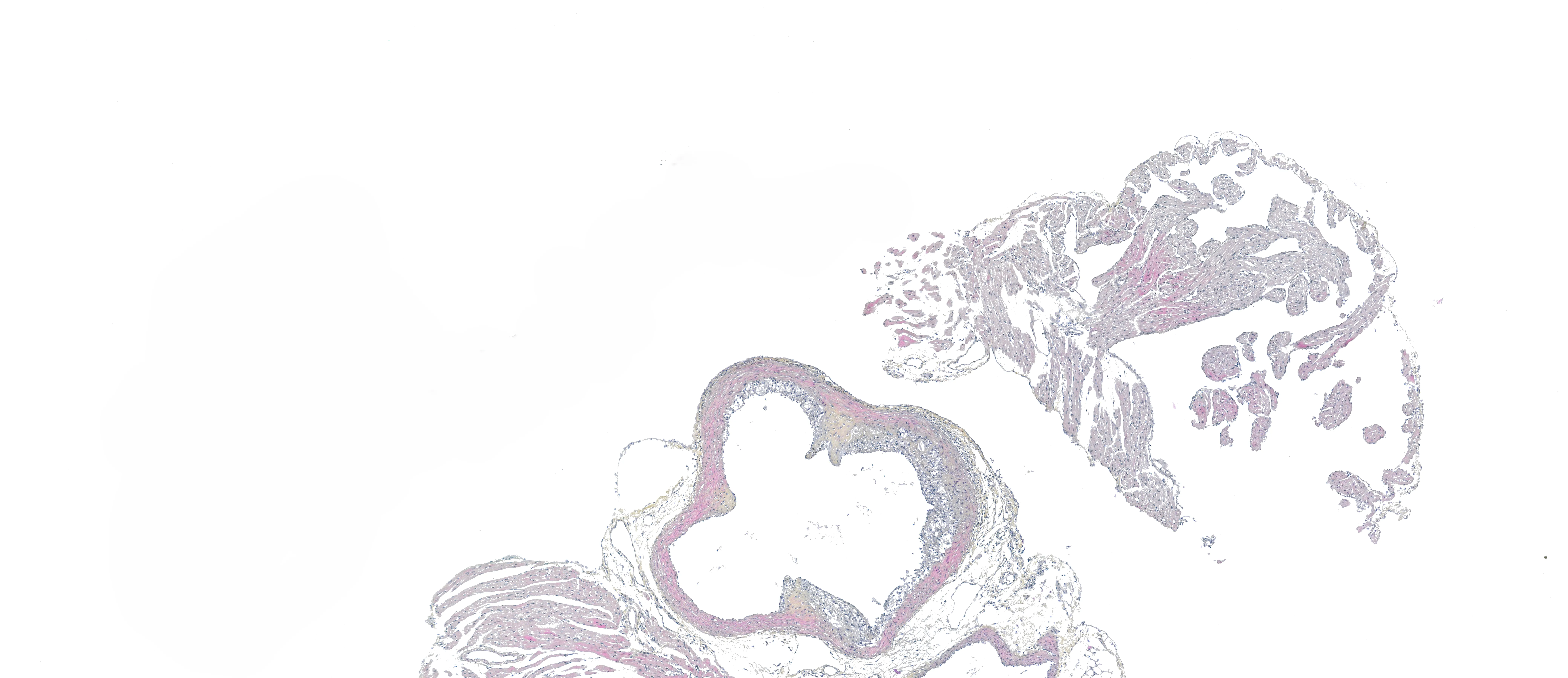

Our digital pathology AI solutions
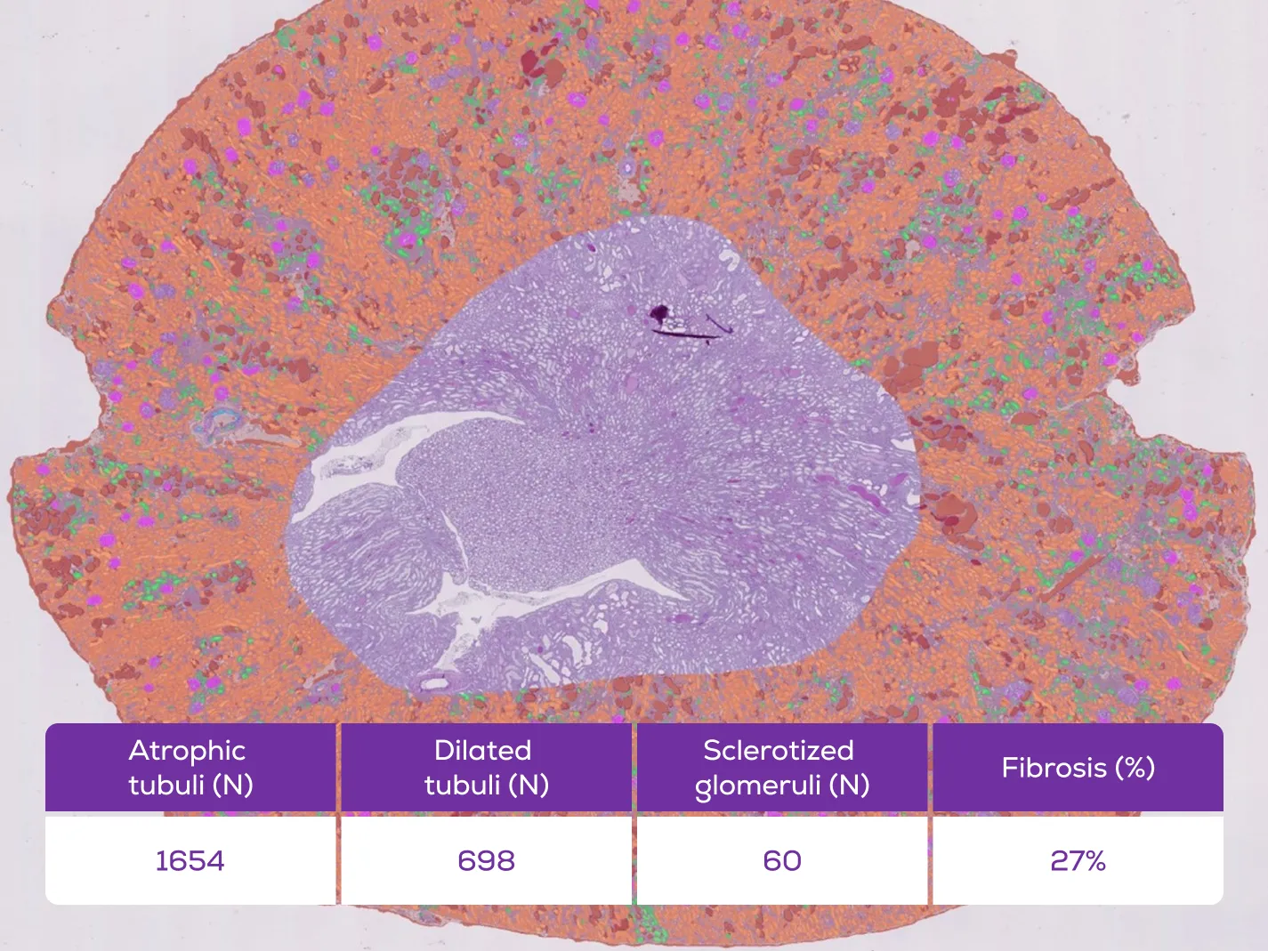
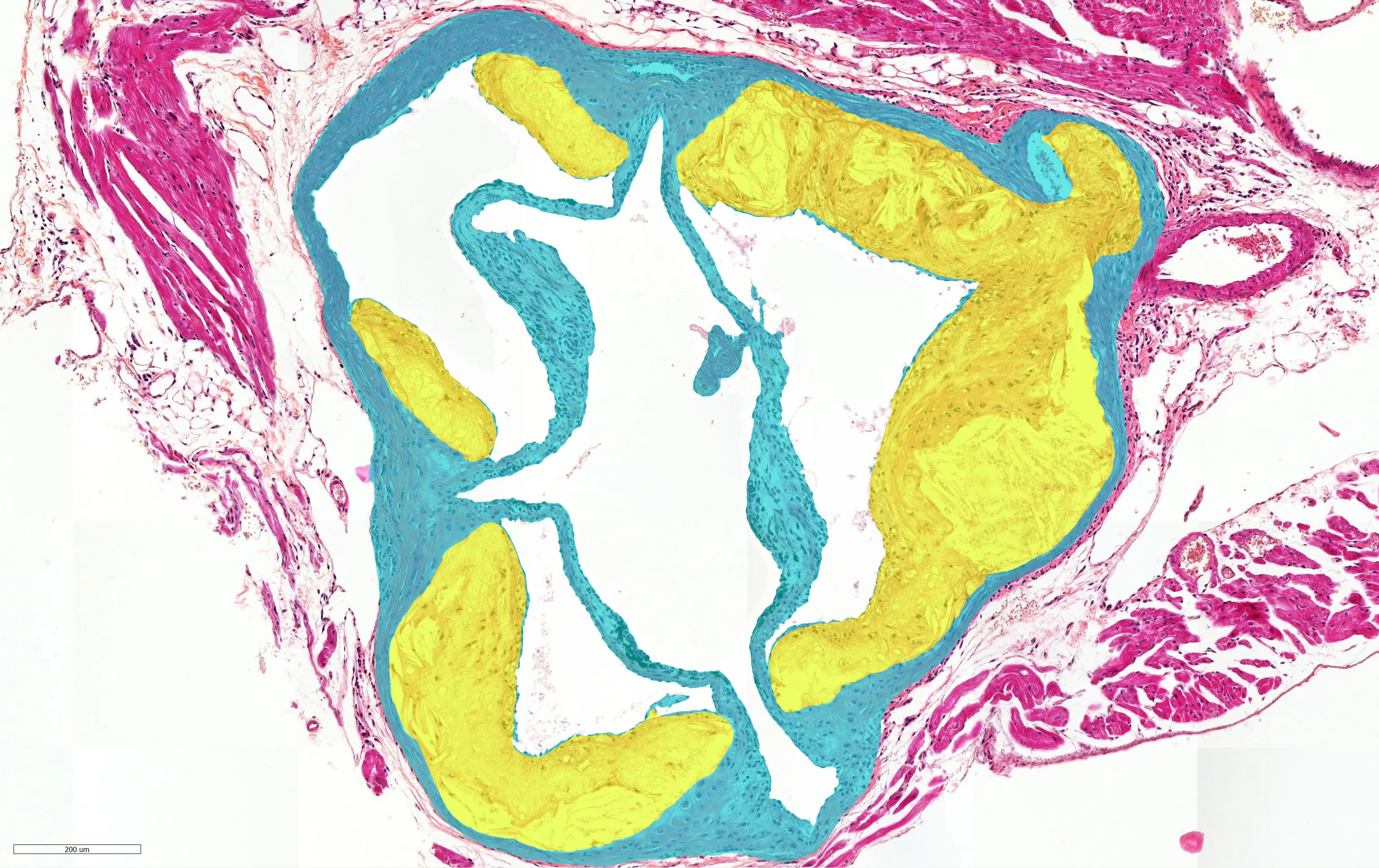
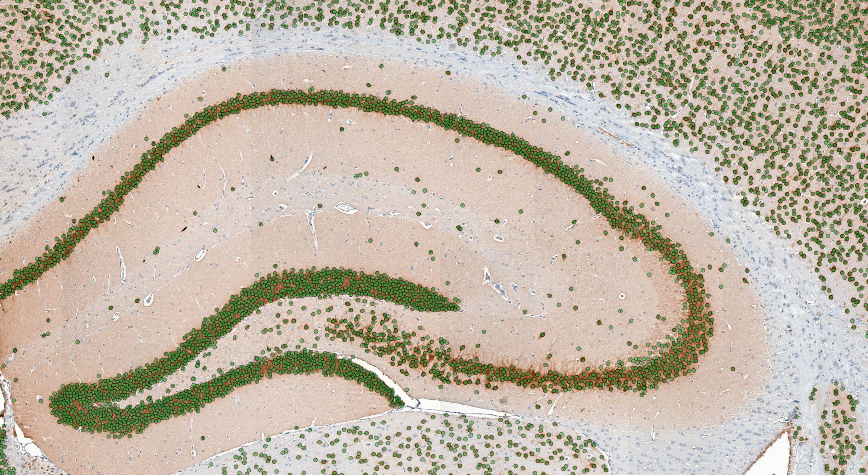
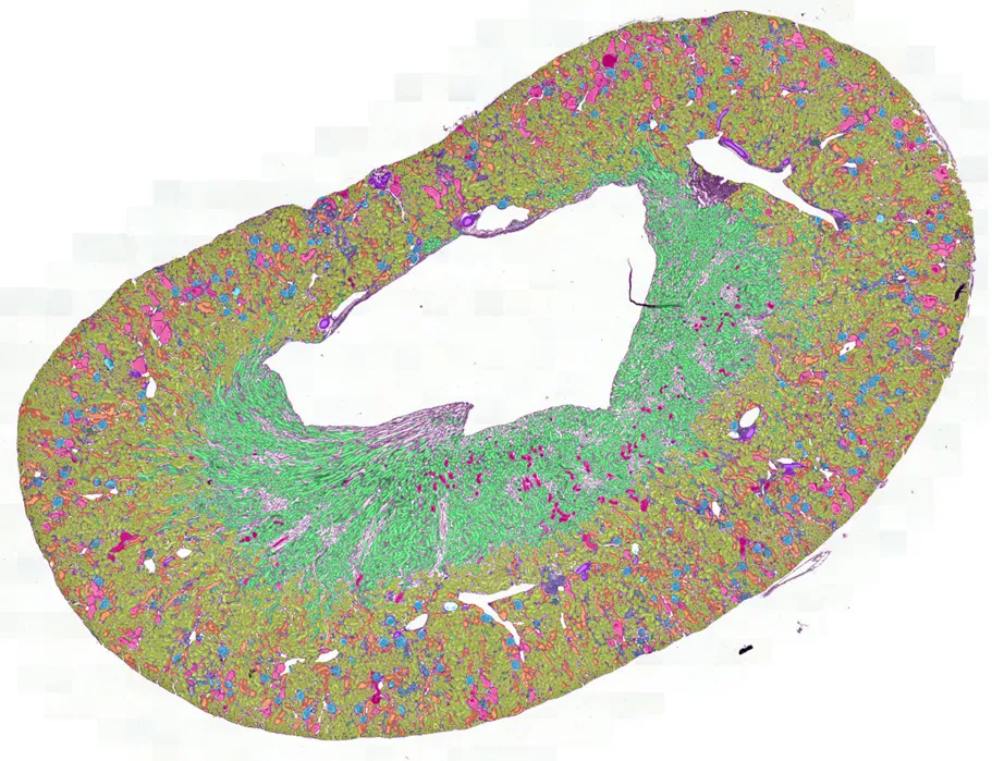
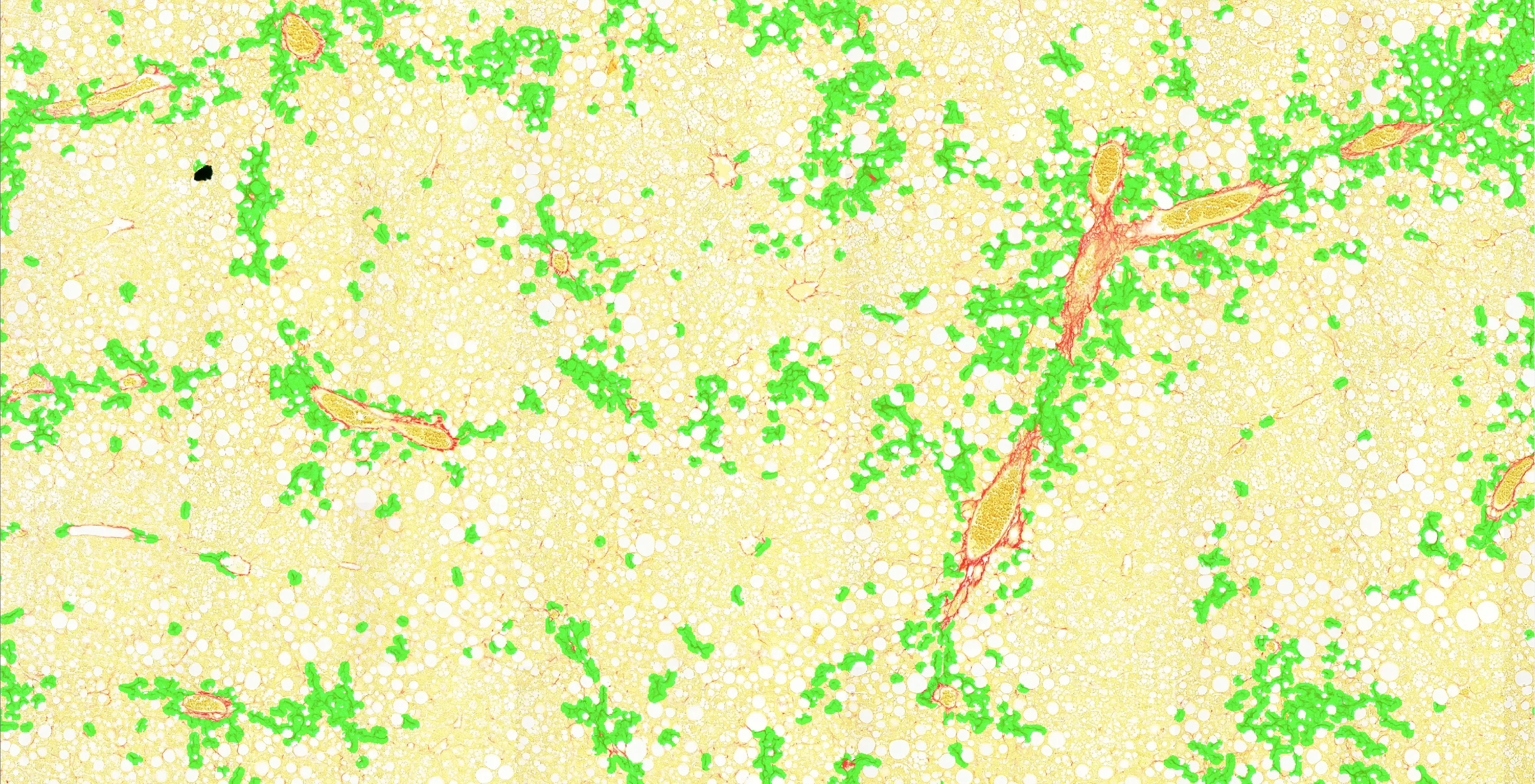
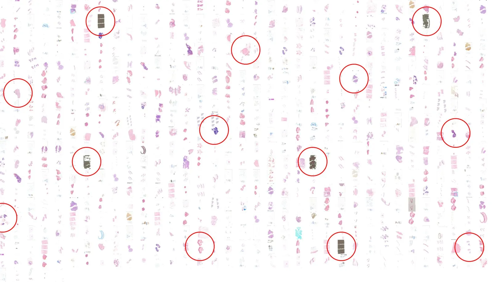
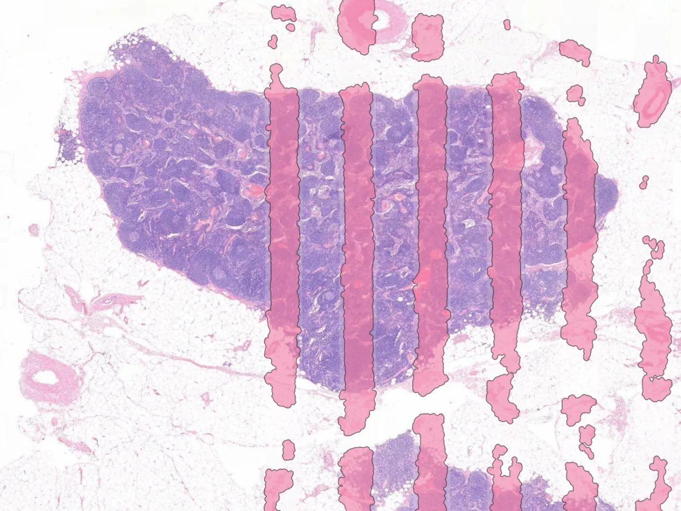
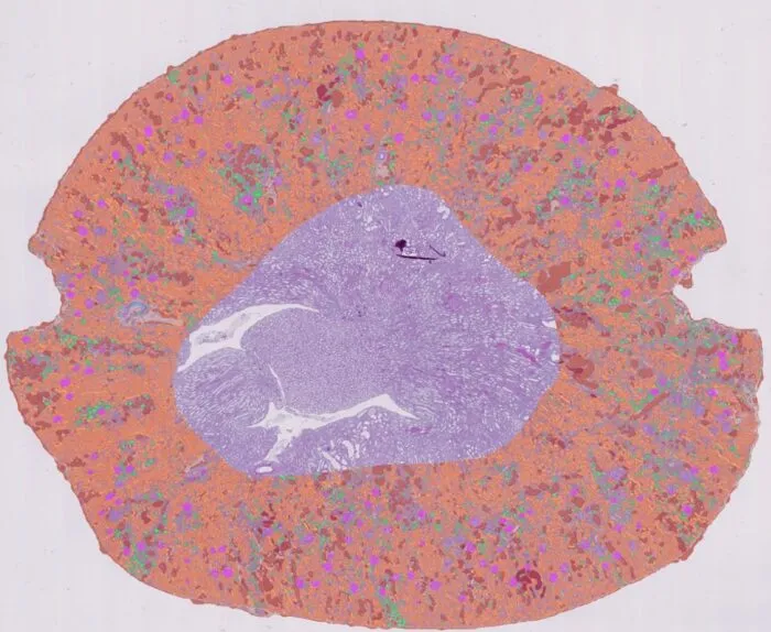
Manual reading samples the section, varies between readers, and does not scale to a full cohort or a full worklist. Our AI brings consistency, so the same slide returns the same read-out, quantified across 100% of the tissue and reported per group at tissue, slide and study level.
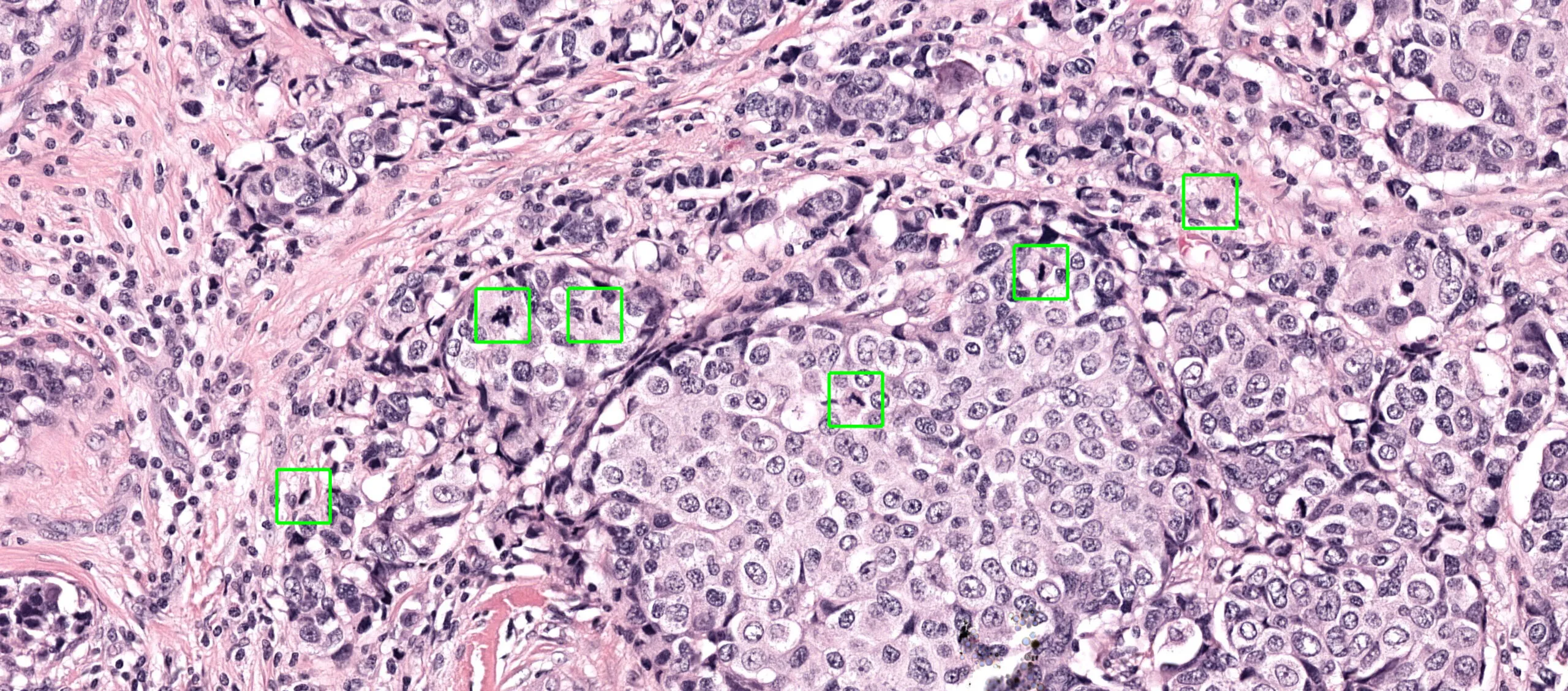
Who we work with
We develop and validate our models with clinical and preclinical partners, and deliver them through the scanner and platform vendors laboratories already use.








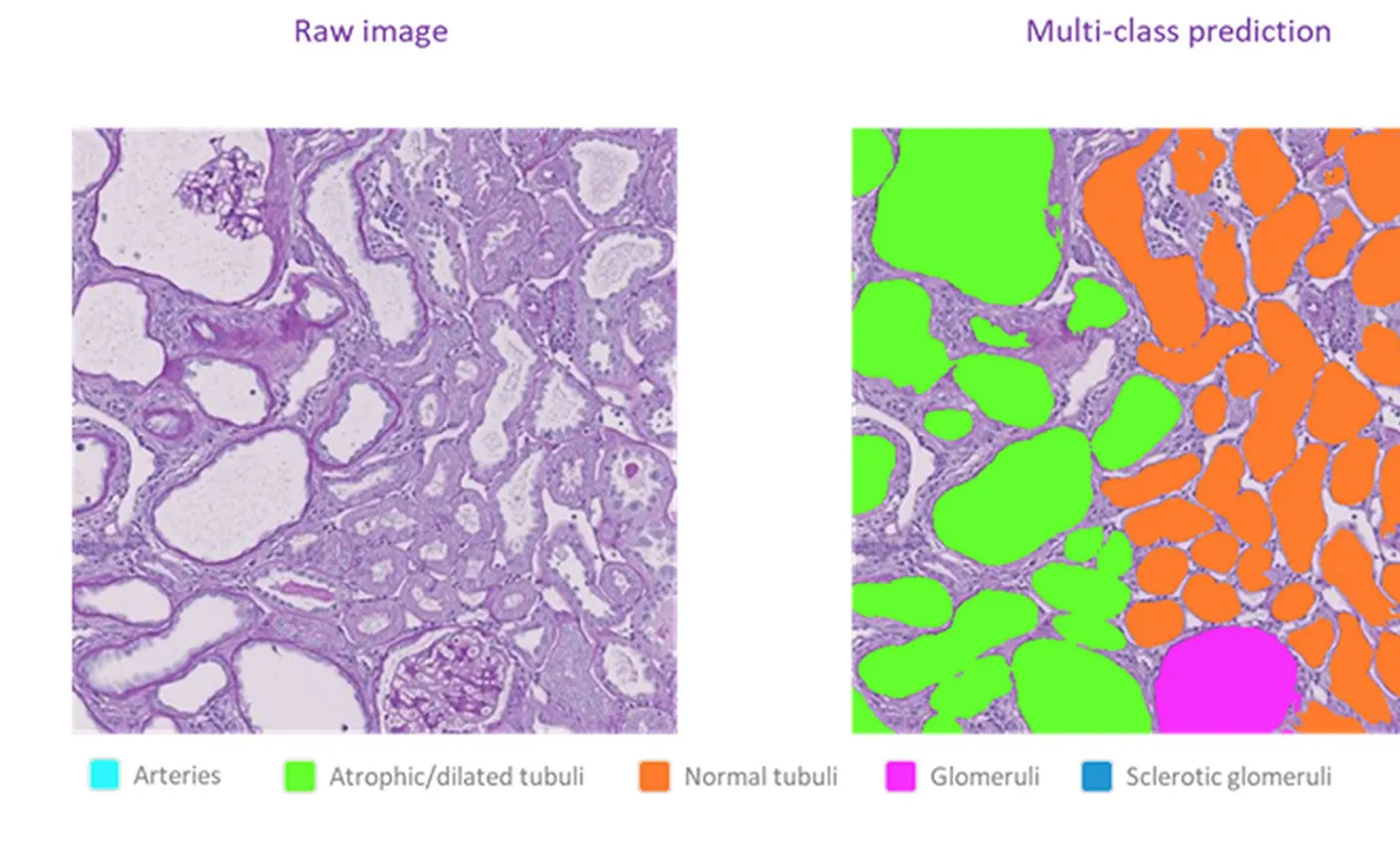
Our AI algorithms applied to the real world
Case studies and examples of our AI solutions used by our partners and customers.
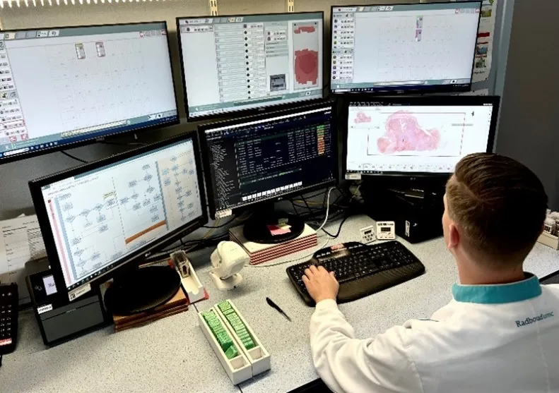
Book a demo of our AI solutions
See the analysis run on whole-slide images and the read-outs it returns. Tell us which area you work in and we will set up a call.
Message sent
Thank you. We will get back to you.