
Objective tissue read-outs for yourpreclinical study
Aiosyn’s computational pathology solutions provide CROs and biopharma organizations with cutting-edge tools to identify new and existing pathology-based biomarkers, enhancing biological understanding and streamlining drug development efforts.
- Every structure on the section, not a sampled field
- Same slide, same read-out, every run
- Validated with preclinical CROs and pathologists
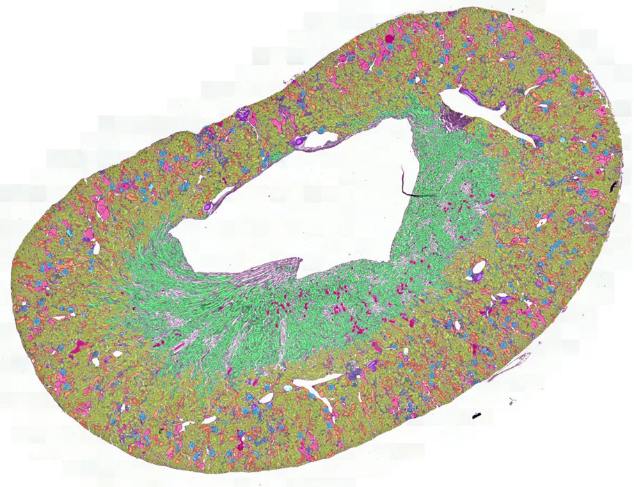
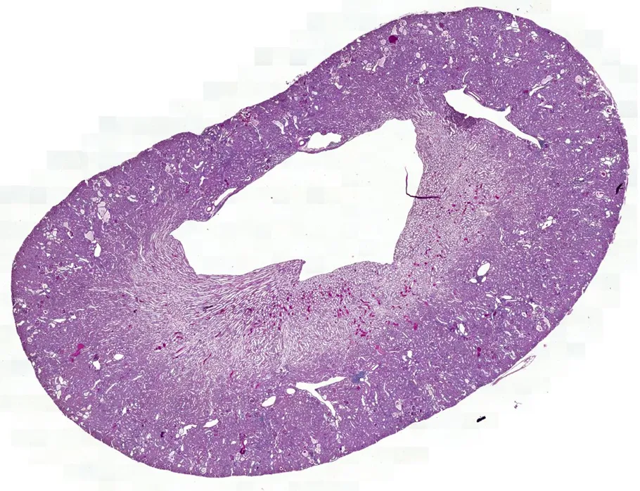 Stained sectionAI read-out
Bringing a new drug to market takes 10 to 15 years
Bringing a new drug to market typically takes between 10 and 15 years and on average $1.3 billion. However, the emergence of transformational technologies such as artificial intelligence and computer vision offers the potential to simplify and accelerate the drug development process, transforming how biomarkers are discovered and developed.
Tissue measured as continuous quantities
Aiosyn’s computational pathology solutions provide CROs and biopharma organizations with cutting-edge tools to identify new and existing pathology-based biomarkers, enhancing biological understanding and streamlining drug development efforts.
Our current focus areas
We build AI solutions for various applications, based and validated on real preclinical cohorts. Explore our focus areas below, or reach out to discuss a different tissue or endpoint.
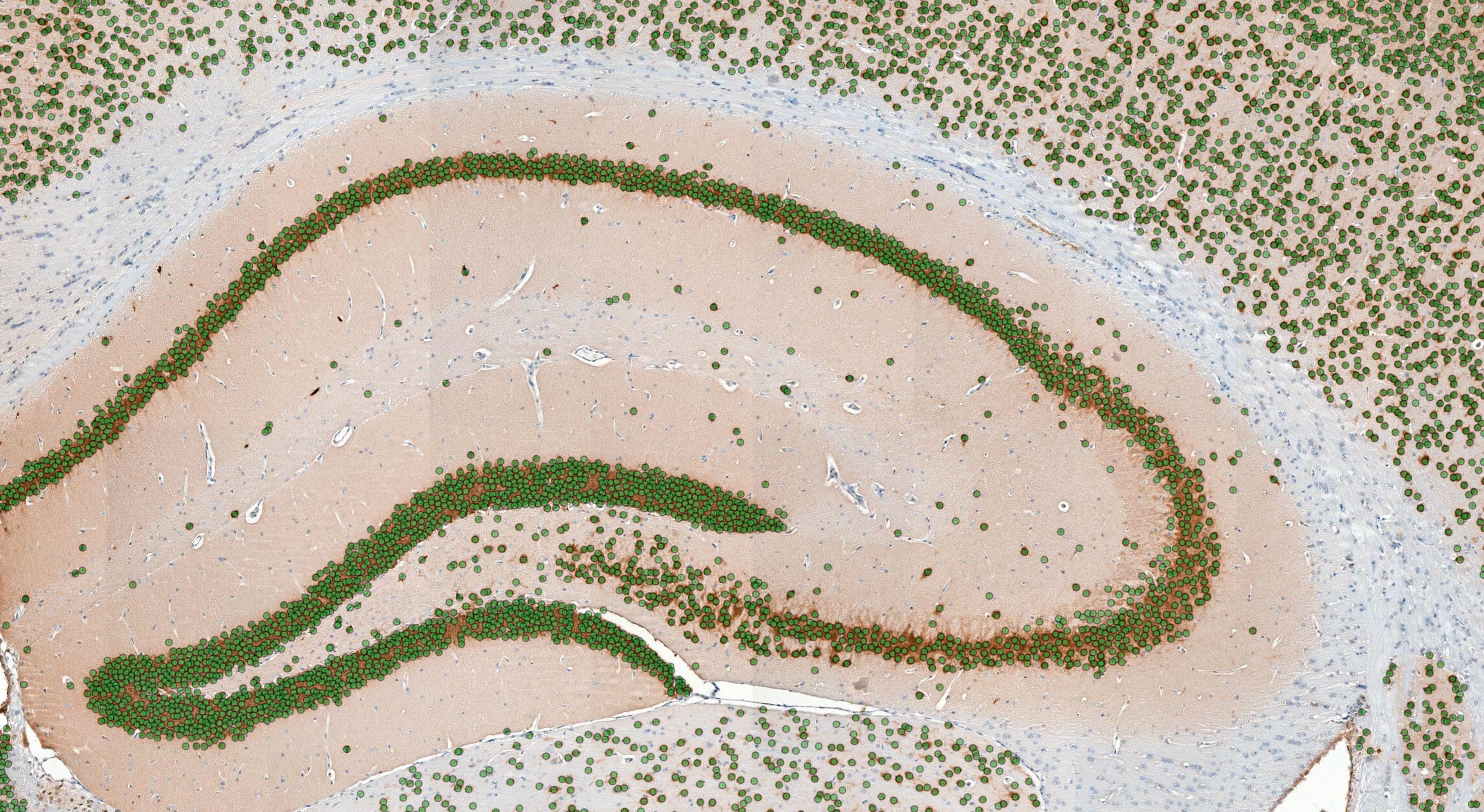
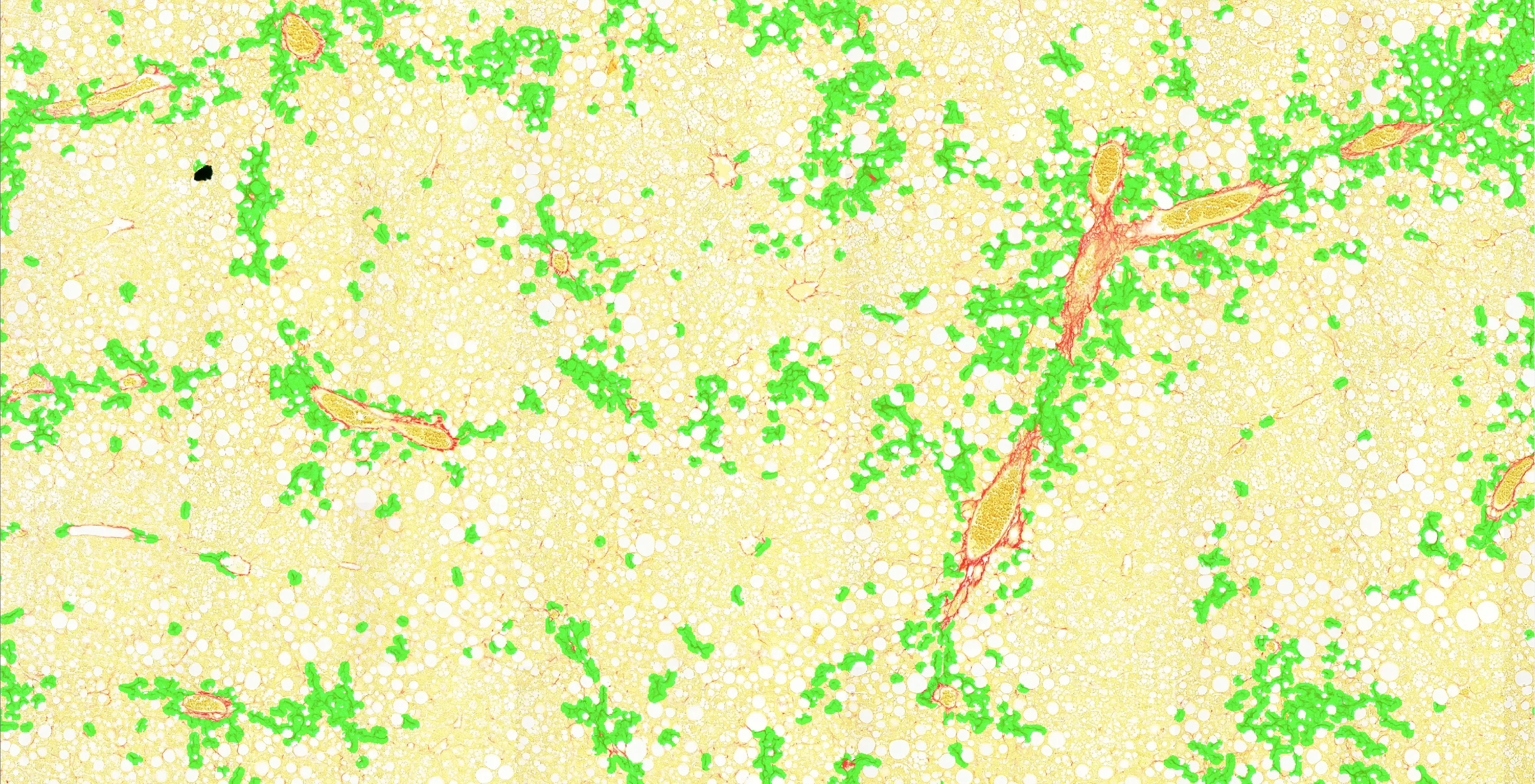

A different tissue or endpoint
Custom AI development builds the model on your own images, for a tissue, stain or endpoint of your interest.
More tissue measured, more statistical power
A manual score samples a few fields per section, and varies with the reader.
The same read-out on every run
One fixed method, applied to every section in the cohort.
Across 100% of the tissue
Every structure on the section is quantified rather than a handful of sampled fields, so the same question can be put to a leaner cohort.
Read-outs inside a week
Where an existing model applies to your tissue, a standard suite analysis returns read-outs within a week.
Validated on real preclinical cohorts
We co-develop and validate our models with pathologists and other experts in the field
Peer-reviewed study

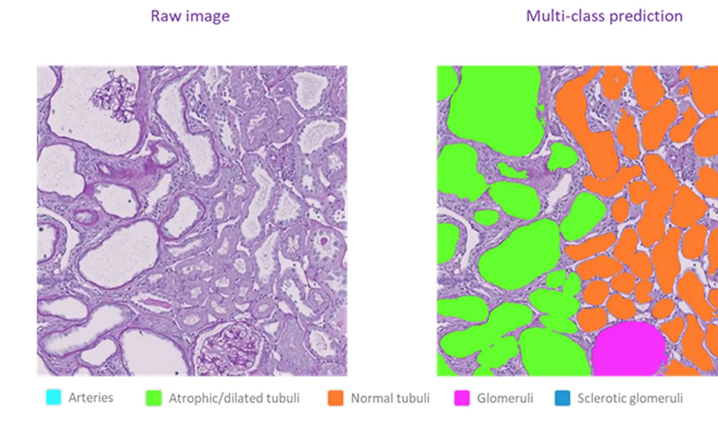
AI read-outs across whole kidney sections detected a treatment effect that manual scoring missed
In a peer-reviewed diabetic nephropathy study with Physiogenex, the AI counted every glomerulus and tubule across the whole kidney section and revealed a dapagliflozin treatment effect.
Briand F, et al. · European Journal of Pharmaceutical Sciences · 2026
Read the study write-upMethod validation
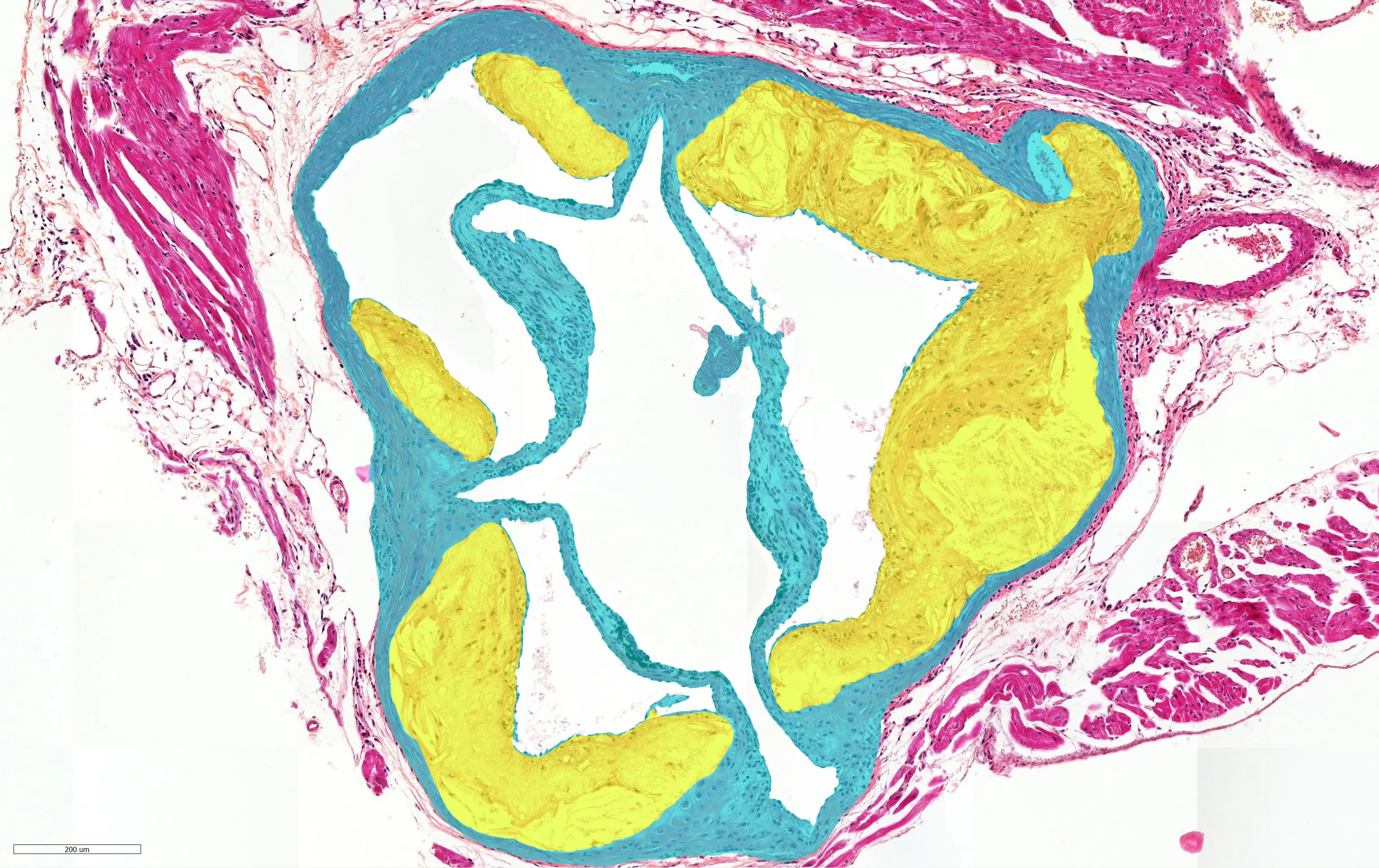
Across 80 cases, AI lesion-area read-outs matched expert pathologist scoring
Developed with TNO, the AI measures atherosclerotic lesion area across the full aortic-root cross-section of every animal in the cohort.
Atherosclerosis · aortic-root cross-sections
Explore atherosclerosis read-outsFrom slide to read-out
Three steps, no new scanning workflow, and fully traceable from case to result.
- 1
Share your slides
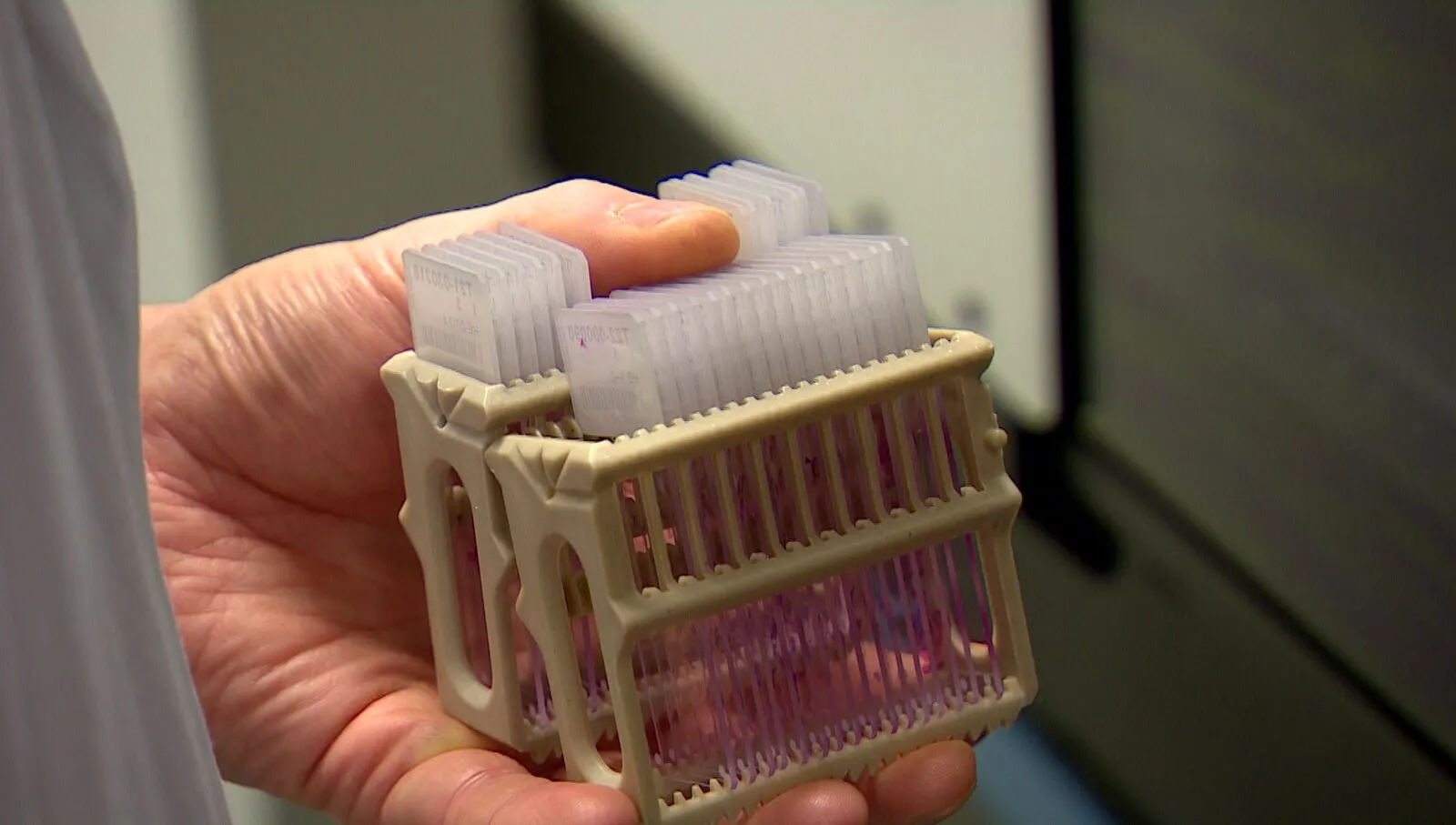
Whole-slide images in the formats you already scan. No change to how your sections are prepared or digitized.
- 2
The AI reads every section
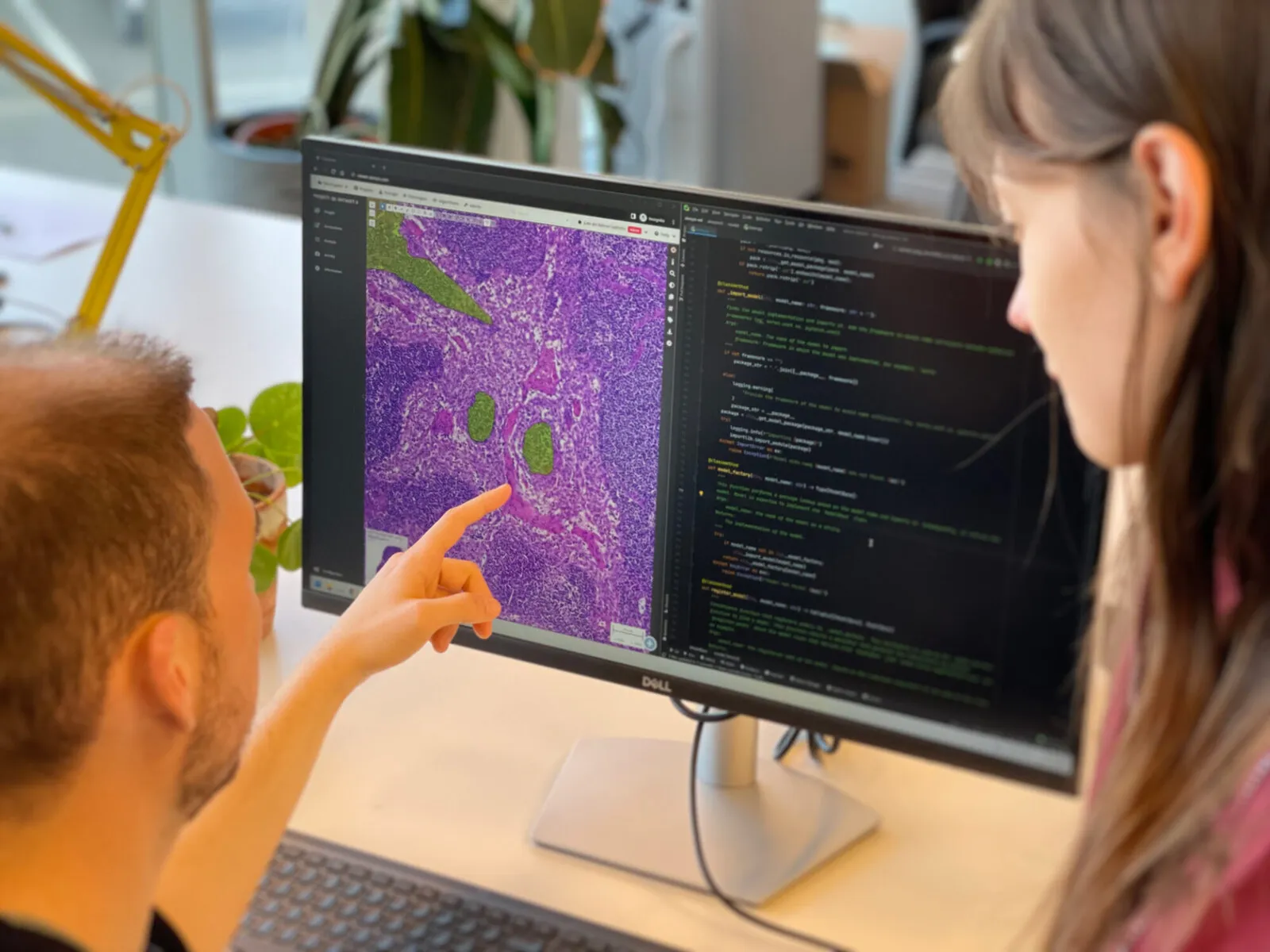
Structures are segmented and cells detected across the full tissue, on every slide in the cohort, under one fixed method.
- 3
Read-outs come back quantified
A report of per-animal and per-group read-outs, ready for your statistics and traceable back to the images they came from.
Let's talk about your cohort
Tell us the tissue, the stain and the endpoint you need measured. We will tell you which read-outs apply and what they would look like on your own sections.