Key takeaways
- Peer-reviewed validation of NephroPath AI: the platform’s whole-kidney quantification of tubular impairment and glomerulosclerosis is now published in the European Journal of Pharmaceutical Sciences, in a study led by Physiogenex with Nipoka, PharmaNest, Sciempath Labo and CLEA Japan.
- Image analysis detected a drug effect that manual scoring missed: classical semi-quantitative histopathological scoring found no positive effect of dapagliflozin on tubuli and glomeruli lesions. NephroPath AI resolved a 32% reduction in dilated tubuli and a 43% reduction in sclerotic glomeruli.
- Counts, not scores: the platform returned absolute numbers of sclerotic glomeruli and atrophic and dilated tubuli across the entire cortical region of each kidney section, not an ordinal score for the slide.
- First application in a rat model: this is the first published use of NephroPath AI in rat kidney tissue, extending the read-outs used in kidney transplant pathology to preclinical efficacy studies.
The study
Diabetic nephropathy is the most frequent complication of type 2 diabetes and the leading cause of end-stage renal disease. Preclinical models are needed to evaluate new therapies, but the imaging read-outs used to assess them are usually semi-quantitative scores assigned by eye, which limits both sensitivity and reproducibility.
Physiogenex set out to optimise drug efficacy studies in the Spontaneously Diabetic Torii (SDT) fatty rat by replacing those scores with three quantitative imaging methods: NephroPath AI for glomerulosclerosis and tubular impairment, the Podocyte Exact Morphology Measurement Procedure (PEMP) from Nipoka for podocyte foot process morphology, and PharmaNest’s FibroNest platform for phenotypic fibrosis.
The panel was tested against a known compound. SDT fatty rats on a 0.3% salt diet, with unilateral nephrectomy (Unx) or sham surgery, were treated with the SGLT2 inhibitor dapagliflozin for 10 weeks. Sham-operated SDT fatty rats and Sprague Dawley rats served as controls. Unilateral nephrectomy and salt supplementation aggravate the phenotype, producing the glomerular filtration rate decline, hypertension and tubulointerstitial fibrosis that characterise later-stage disease.
What NephroPath AI measured
NephroPath AI segments and delineates kidney structures on digitised PAS-stained slides. Slides were scanned at 0.5 µm/px and analysed at 1.0 µm/px. The model segmented glomeruli including sclerotic glomeruli, proximal, distal, atrophic and dilated tubuli, arteries, capsule and interstitium across the cortical region, and the total number of sclerotic glomeruli, atrophic tubuli and dilated tubuli per whole kidney section was extracted as a quantitative histopathological endpoint. All image analysis was performed blindly, without any information about treatment groups.
The model tracked disease severity as expected. Atrophic tubuli were 3-fold higher in sham SDT fatty rats than in Sprague Dawley controls and 4-fold higher again in Unx rats (p < 0.01). Sclerotic glomeruli followed the same pattern, 5-fold greater in sham rats and 6-fold higher in Unx rats than in sham (p < 0.0001).
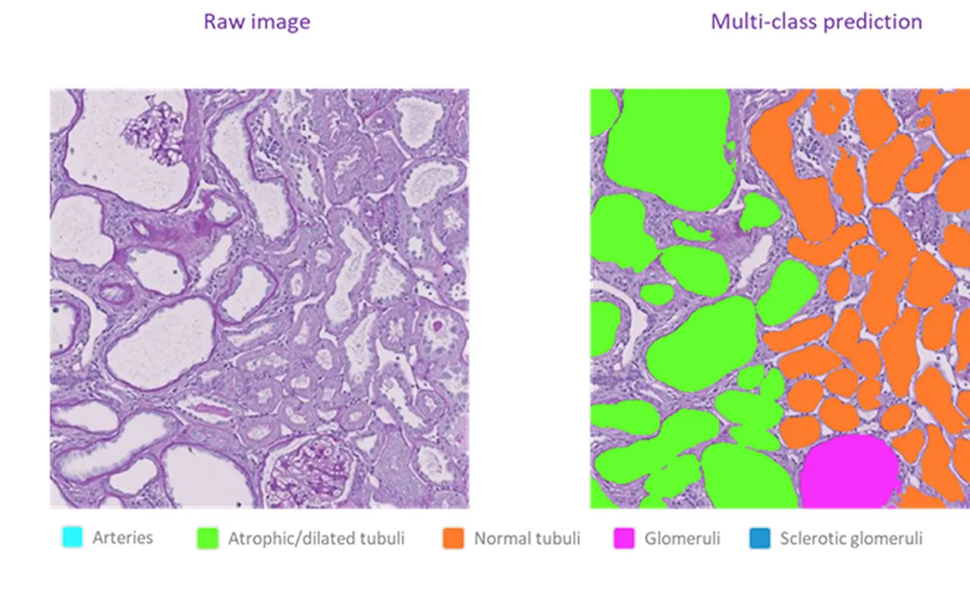
Figure 1. Raw PAS staining and the NephroPath AI multi-class prediction of the same region. Legend: arteries (cyan), atrophic and dilated tubuli (green), normal tubuli (orange), glomeruli (magenta), sclerotic glomeruli (blue).
Where the AI read-outs separated from manual scoring
The comparison the paper makes between methods is the part most relevant to study design. Using the conventional histopathological scoring system, the Unx group showed the expected worsening of tubular atrophy, tubular casts and interstitial fibrosis, but the scores did not indicate any positive effect of dapagliflozin on those lesions. Only the inflammation marker (%ED1 immunostaining) showed a significant reduction.
The quantitative read-outs did resolve an effect. NephroPath AI measured a significant 32% reduction in dilated tubuli and a 43% reduction in sclerotic glomeruli in treated Unx rats (both p < 0.05), with a 19% reduction in atrophic tubuli that did not reach significance. PEMP showed a significant improvement in filtration slit density, indicating recovery from podocyte effacement, and FibroNest showed a significant reduction in the assembled fibrosis morphometric composite score in the cortex.
In other words, the treatment effect was there. The scoring system was not sensitive enough to see it in the tubuli and glomeruli, and counting every structure across the section was.
Why this matters for preclinical studies
A read-out that misses a real effect costs a programme more than a slow one. Semi-quantitative scoring compresses a whole section into a small ordinal scale, and effects of the size seen here fall inside the noise of that scale. Quantifying every glomerulus and tubule instead produces continuous endpoints on the same slides, with no additional tissue and no change to the animal model.
The authors conclude that this image analysis represents a good alternative to classical semi-quantitative analysis in PAS-stained kidney sections for detecting renoprotective effects of therapies in animal models, and that the techniques are applicable in both animals and humans, which supports translation from preclinical work to the clinic.
This publication builds on the earlier collaboration with Physiogenex described in our case study on quantitative assessments of kidney health in diabetic animal models.
Publication details
Briand F, Grasset E, Shinohara M, Cordeiro L, Richard V, Endlich N, Endlich T, Chen L, Petitjean M, de Bel T, Sulpice T.
European Journal of Pharmaceutical Sciences, volume 224 (2026), 107588. Available online 17 June 2026. Open access under CC BY-NC-ND 4.0.
- The NephroPath platform is for Research Use Only and should not be used for diagnostic procedures.