Nijmegen, the Netherlands – 1 September 2026. Aiosyn has launched its new website that reflects its current focus: quantitative AI-based tissue analysis for preclinical studies and automated quality control for clinical laboratories. The new structure highlights Aiosyn’s sharpened product offering for biopharma and clinical laboratories following its 2025 funding round and strategic partnership with TNO.
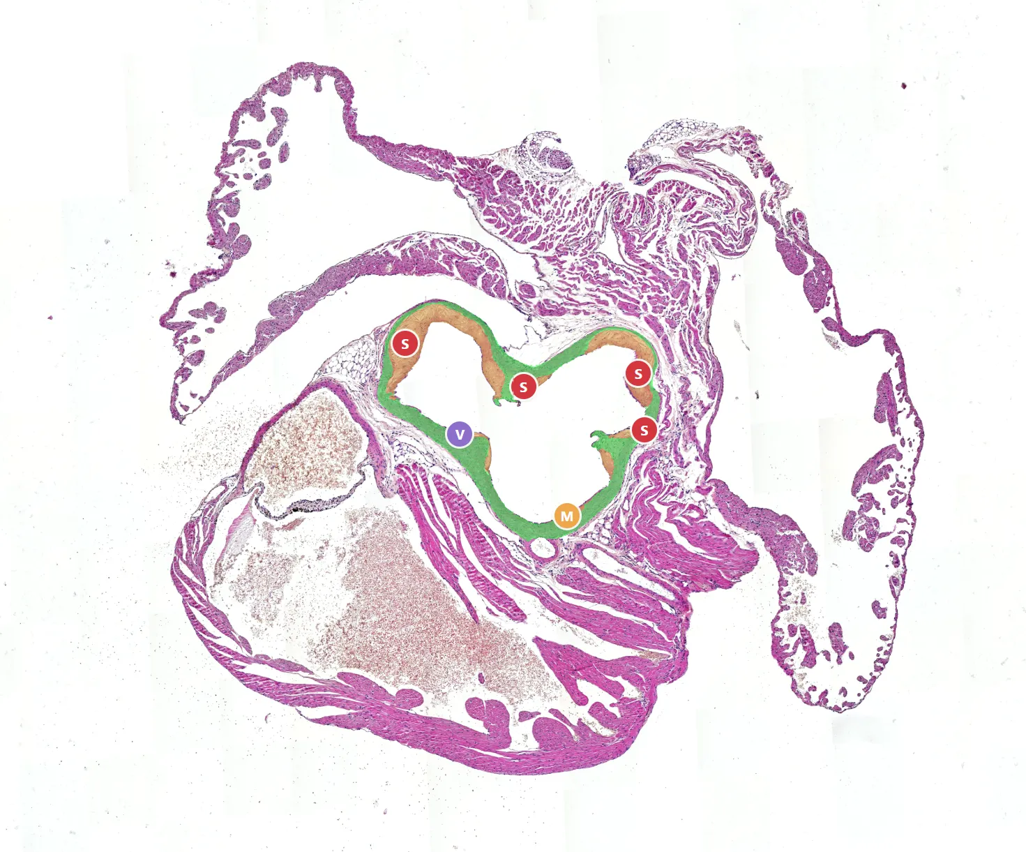
Aiosyn’s work does not start with AI. It starts with tissue and the questions that tissue can answer. Is there a treatment effect on atherosclerotic lesions across this cohort? Is this kidney tissue deteriorating? Is this digital slide ready for pathologist review, or should it return to the laboratory? Aiosyn’s new website is organized around these questions, and how they can bring value to biopharma and clinical laboratories.
Preclinical studies
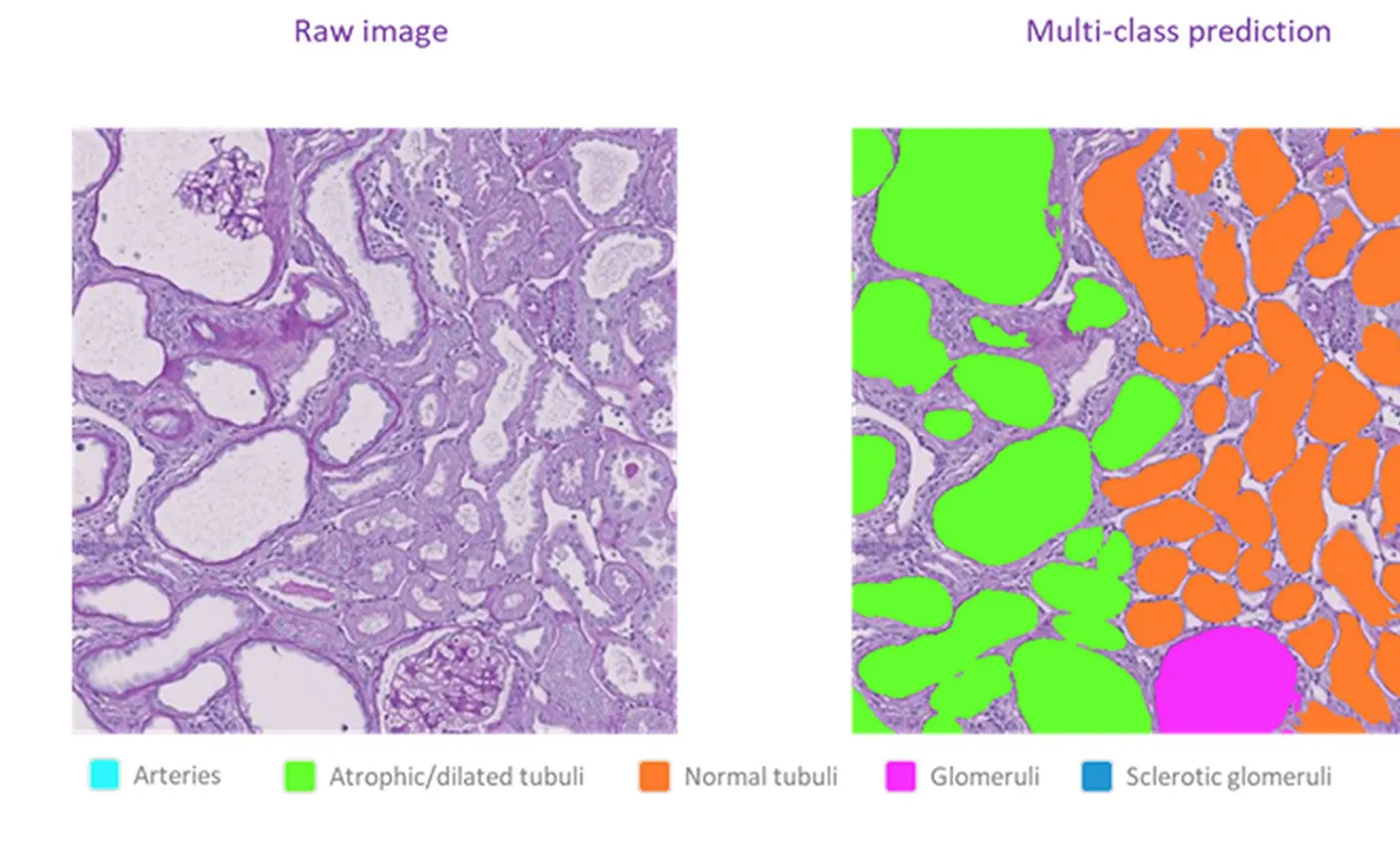
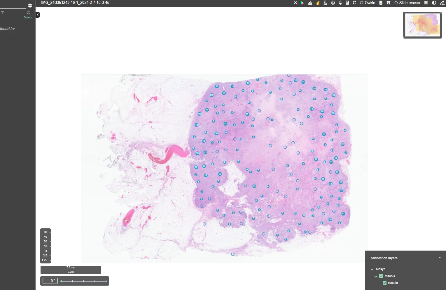
Histological assessment is a common endpoint in many drug-development studies. Aiosyn’s image analysis measures structures and changes across whole-slide images, returning quantitative read-outs per animal that can be traced back to the underlying tissue.
Aiosyn’s biopharma work includes new models for atherosclerotic lesions and kidney, brain and liver tissue, developed together with TNO. Combined, they form the foundation of a multi-organ platform that Aiosyn is building to cover the major organs assessed in preclinical studies.
Clinical laboratory workflows
For clinical laboratories, the site introduces new features for AiosynQC within the context of the full digital pathology workflow.
AiosynQC checks digitized H&E and IHC slides for common image-quality problems, including out-of-focus regions, tissue folds, incomplete scans and white-balance errors. It supports laboratory teams in deciding whether a slide is ready for pathologist review or requires rescanning or new preparation, based on the laboratory’s own acceptance criteria. Its dashboard brings QC results together, helping laboratories identify recurring issues and make data-driven improvements to their digital pathology workflow. AiosynQC integrates with the scanners, image-management systems and viewing platforms laboratories already use.
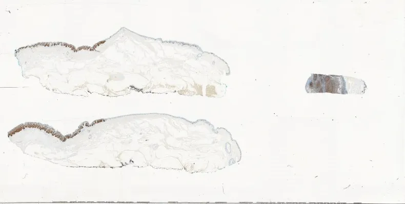 Macro image of the glass slide
 Whole-slide image
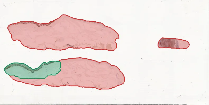 AiosynQC read-out
Missing from the whole-slide imageCaptured
- Tissue missing
- 88%
- Tissue captured
- 12%
About Aiosyn
Aiosyn develops computational pathology software for preclinical studies and clinical laboratory workflows. Its solutions turn whole-slide images into quantitative tissue read-outs and automate selected steps in digital pathology workflows.