
Automated quality controlfor digital pathology slides
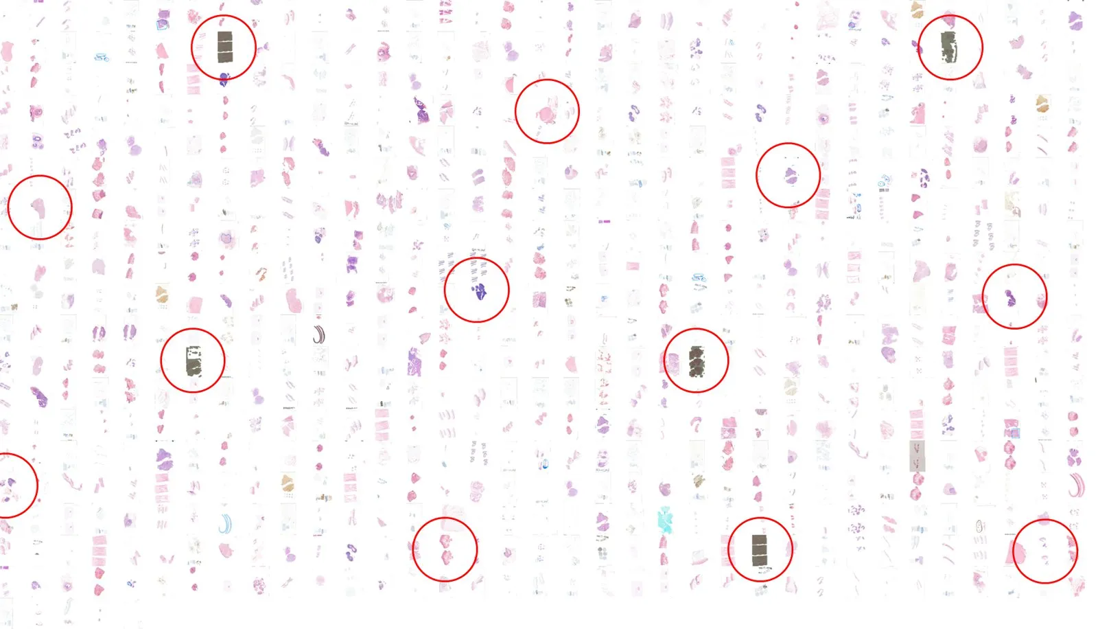
Variation in slide preparation and scanning leaves whole-slide images that are hard to read — or, incomplete, with tissue missing from the scan. Found late, either problem means a rescan after the case has reached a pathologist. AiosynQC checks every image as it arrives, segments the affected or missing regions, and flags the slides that need a new preparation or a rescan.
In the EU and the UK, AiosynQC is not a medical device under IVDR and UK MDR 2002. Research Use Only in the United States.
- Every whole-slide image checked, at pixel level
- H&E and IHC, on-premise or in the cloud
- Results in the platform you already work in
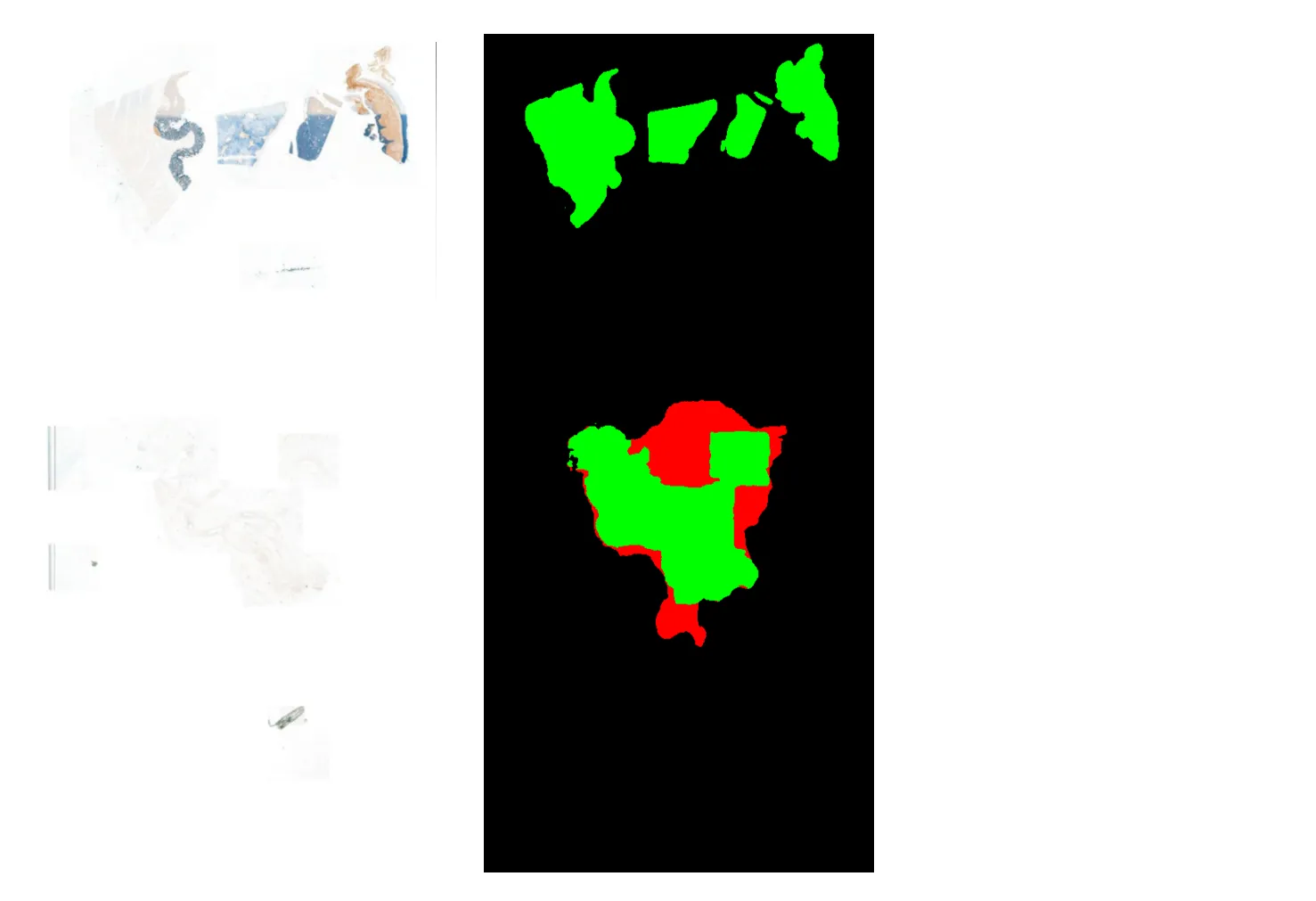
Every affected region marked on the image itself
Drag the handle across one H&E section. This scan picked up out-of-focus banding, so the flagged slide arrives with the location and extent of the problem rather than a pass or fail verdict.
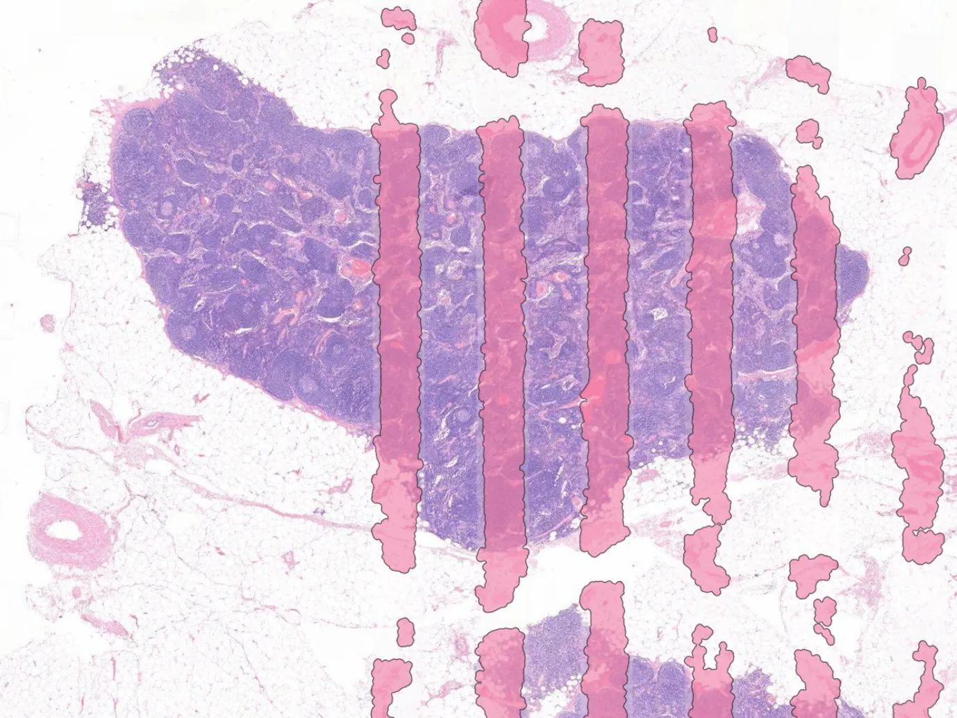
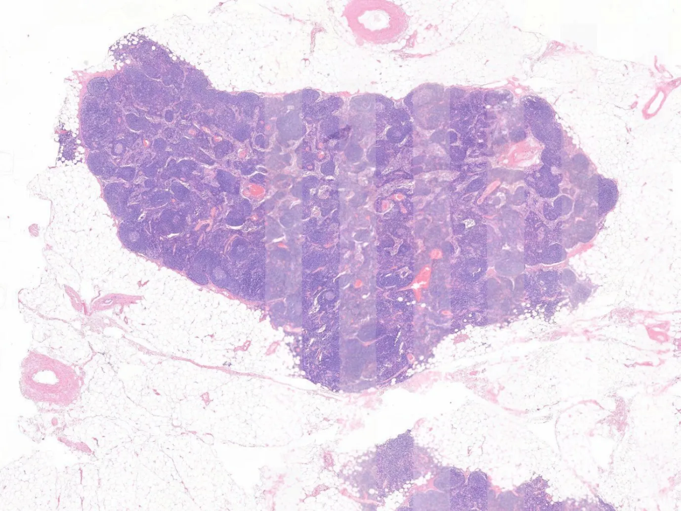 H&E sectionDetected artifacts
Every common artifact, on H&E and IHC
The algorithm classifies every slide against your laboratory's acceptance criteria. Sensitivity is set per laboratory, so reporting can flag tissue folds above a size threshold while smaller ones pass.
- Out-of-focus areas
- Incomplete scanning
- Air bubbles
- Tissue folds
- Pen markers
- Dust
- White balance problems
- Ink
Your workflow with AiosynQC
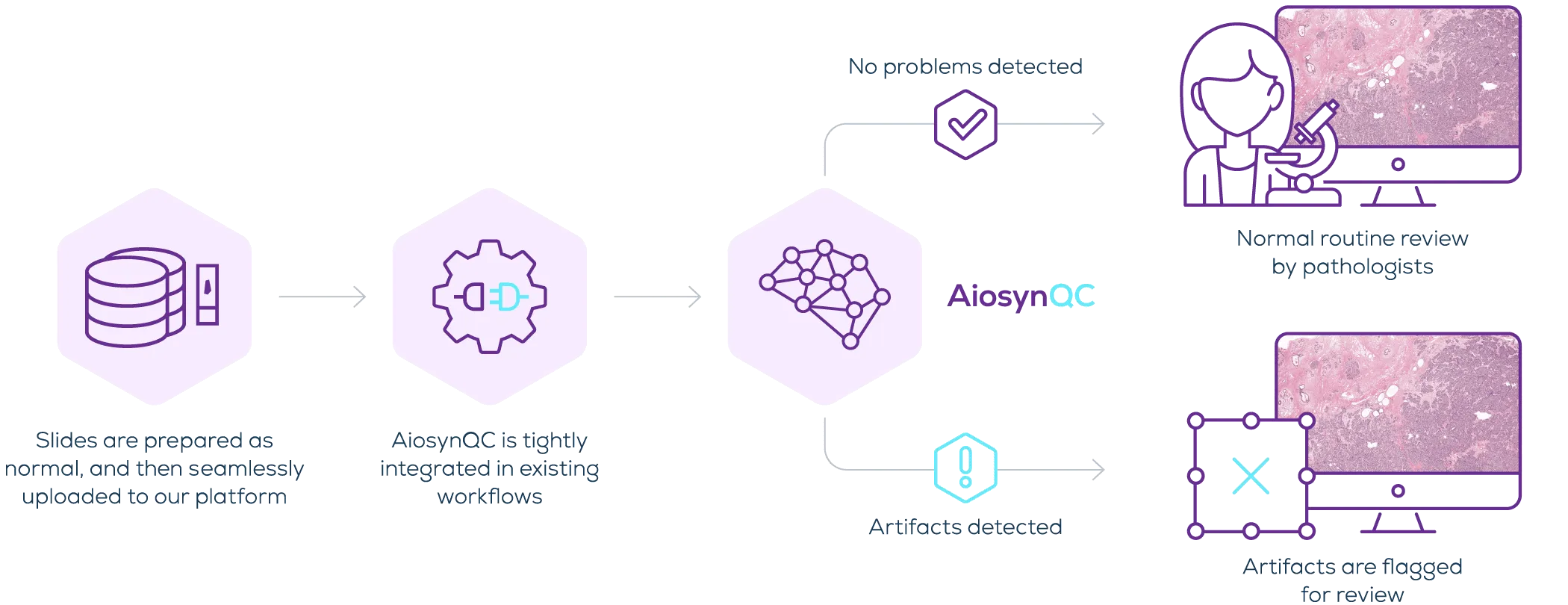
Nothing changes in the laboratory or for the pathologist. Slides are prepared and scanned as usual, every image is analyzed as it arrives, and only the flagged ones need anyone's attention.
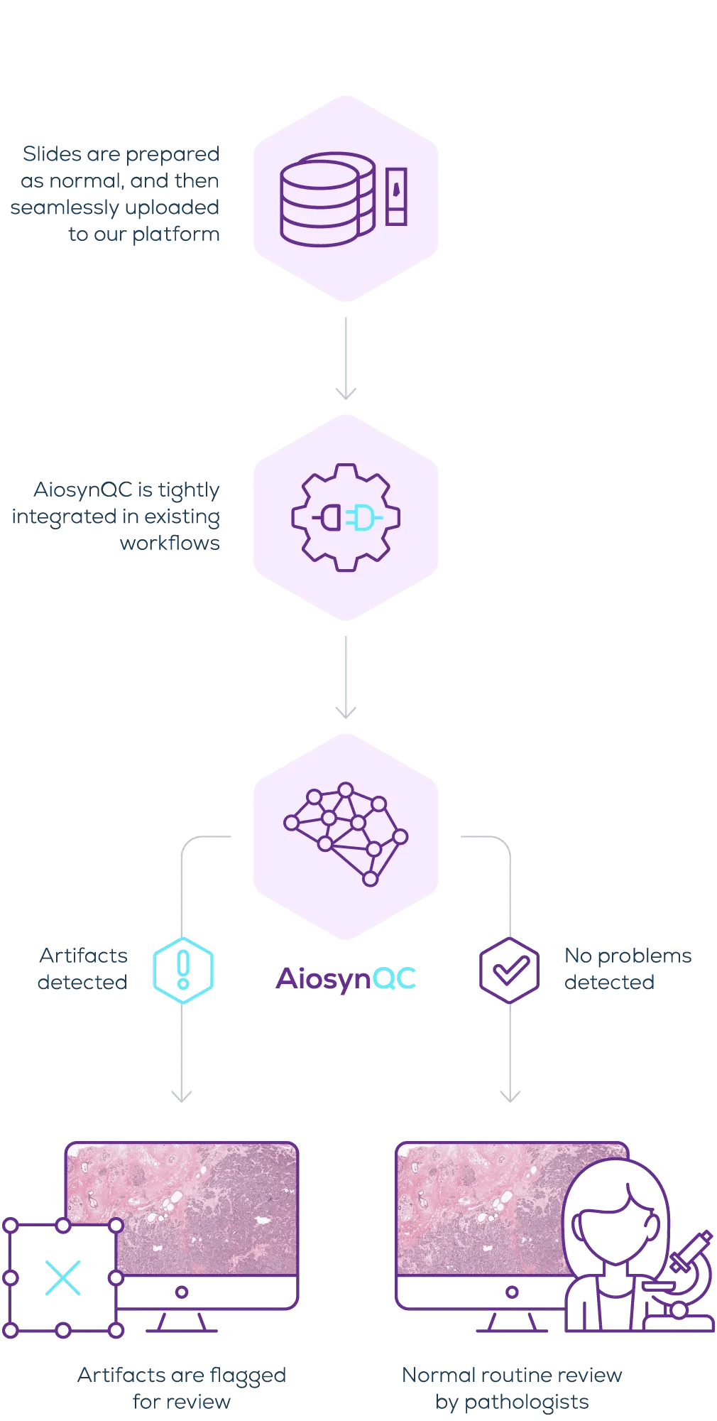
Integration and deployment
Available through Sectra Amplifier Marketplace, integrated in platforms such as Techcyte Fusion, or connected directly to your image management system. On-premise, inside your own network, or in the cloud.
What automating the check changes

Fewer unreadable slides reach a pathologist
Artifacts introduced in preparation or scanning are caught at the pre-analytical step instead of during diagnosis, so a rescan does not cost a diagnostic delay.

Hours of manual inspection you do not have to spend
Out-of-focus regions and tissue folds are rarely visible at low magnification, so checking for them by hand means opening and zooming into every slide — and checking for incomplete scanning means comparing that image back against the physical glass slide.

Clean inputs for the algorithms that follow
Downstream AI underperforms on slides with artifacts. Automated quality control makes the inputs to other algorithms, and to model training, predictable.
I am excited to see AiosynQC fully integrated and seamlessly supporting our lab technicians. It is a great help to catch artifact-affected slides before they reach our pathologists without requiring zoom-ins to manually inspect the images.
Head of the Department of Pathology

The most frequent questions about AiosynQC
Is AiosynQC limited to hematoxylin and eosin (H&E) staining, or can it assess immunohistochemistry (IHC) slides as well?
AiosynQC isn't limited to H&E — it also supports IHC and other special stains.
Does the tool only classify a slide as having artifacts, or does it also segment the affected area?
AiosynQC detects the most common quality artifacts and segments the affected areas.
What types of artifacts can the software detect?
AiosynQC can detect the most common quality artifacts in whole-slide images, including out-of-focus areas, incomplete scanning, air bubbles, tissue folds, pen markers, dust, white balance problems, and ink.
How does the output of your models look, and how can it be customized to specific workflows?
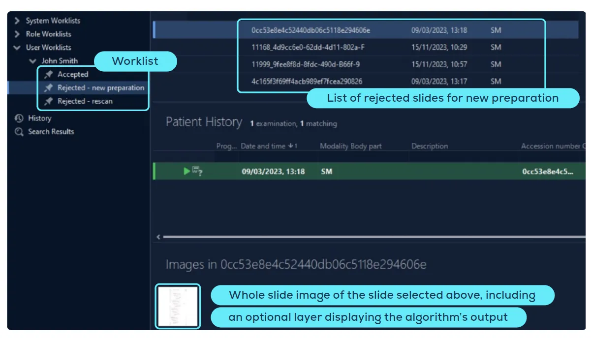
AiosynQC can provide results in multiple formats, including worklists of accepted and rejected slides, detailed quality scores, and color-coded indicators that highlight the presence or absence of artifacts. For a more in-depth analysis, users can click on a whole-slide image to view an overlay that segments affected regions and indicates the artifact types. The output and reporting of compromised slides can be tailored to meet the specific needs of the laboratory. For example, labs can configure the system to flag only tissue folds exceeding a certain size threshold while ignoring smaller ones.
What file types are compatible with AiosynQC?
The tool supports DICOM, TIFF, and OpenSlide-compatible formats such as SVS, MRXS, and more.
Can AiosynQC be run on-premise?
Yes, the software can be run on-premise as well as in the cloud.
Is the software CE-marked?
AiosynQC is not CE-marked because, under the European IVDR, it is not considered a medical device in the EU, nor is it intended to be used as an accessory to any AI or other medical devices.
What percentage of slides are flagged by AiosynQC?
The incidence of artifacts varies depending on the laboratory workflow and equipment. AiosynQC offers tailored sensitivity and customized reporting options to adapt to the specific needs of each laboratory. For instance, labs may choose to flag images with significant anomalies while ignoring small artifacts that typically do not compromise readability. Other centers may prefer a more comprehensive review, to ensure that all images sent to the pathologist are of high quality and do not require a rescan.
Quality control in the news
Also in the clinical track
Clinical overview- AiosynQCAutomated whole-slide quality control
- Mitosis (breast)Mitotic figure counting for breast cancer
- Custom AI DevelopmentA model built for your tissue, stain or endpoint
See AiosynQC on your own slides
Send us a set of whole-slide images and we will show you what the algorithm flags, and how the result would land in your workflow.