
Every image checked,every artifact located
Variations in preparation and scanning leave artifacts and missed tissue areas that are not visible at the scale a collection is reviewed. AiosynQC analyzes every whole-slide image at pixel level, segments the regions they affect, and returns the result as data.
In the EU and the UK, AiosynQC is not a medical device under IVDR and UK MDR 2002. Research Use Only in the United States.
- H&E, IHC, and other special stains
- DICOM, TIFF, and OpenSlide-compatible formats such as SVS, MRXS, and more
- Sensitivity set per collection
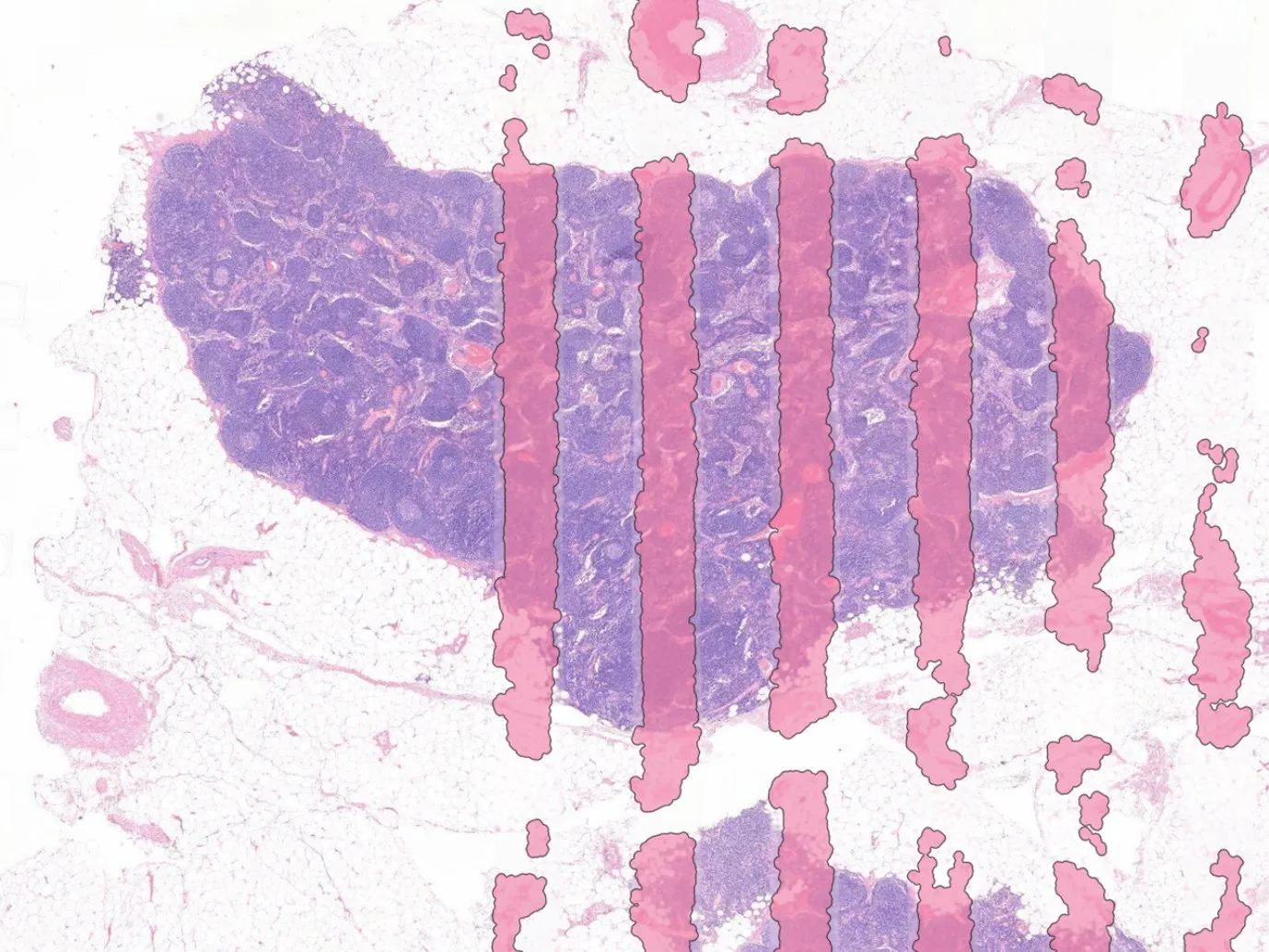
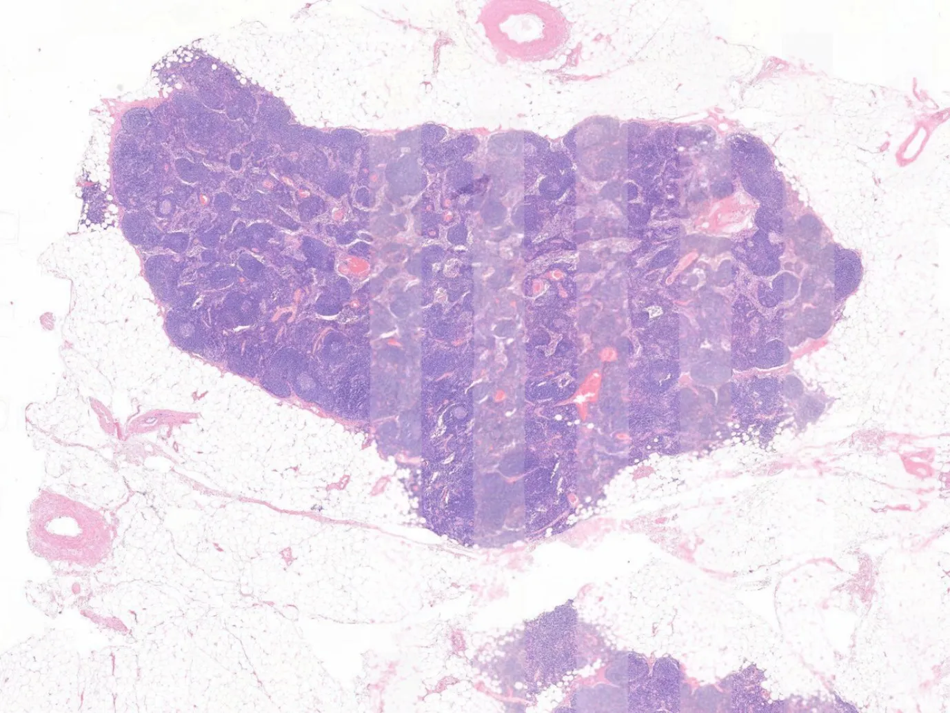 As scannedDetected artifacts
Analysis runs where the images already are
AiosynQC reads from the storage the collection already lives in, on-premise or in the cloud. Nothing is re-scanned.
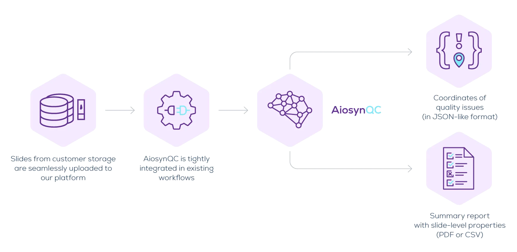
One output filters the collection, the other masks the tile
The slide-level summary
Every image in the collection with the artifact types found on it and the share of tissue they cover, as PDF or CSV.
| slide-00415 | clean | 0.0% |
| slide-00417 | out of focus, pen marker | 3.8% |
| slide-00418 | incomplete scan | 19.2% |
| slide-00421 | tissue fold | 1.2% |
The artifact coordinates
Each region the algorithm segments is returned as geometry, in a JSON-like format, with its type and the area it covers.
{
  "slide": "slide-00417",
  "findings": [
    { "type": "out_of_focus",
      "area_mm2": 12.4,
      "polygon": [[8214, 3390], ...] },
    { "type": "pen_marker",
      "area_mm2": 2.1,
      "polygon": [[1180, 9042], ...] }
  ]
}
Both panels illustrate the shape of the output, not real results.
Every common artifact, on H&E and IHC
The algorithm classifies the slide against the thresholds set for the collection and segments the regions it flags.
- Out-of-focus areas
- Incomplete scanning
- Air bubbles
- Tissue folds
- Pen markers
- Dust
- White balance problems
- Ink

Streamline slide review processes
Automate image quality control with AiosynQC to reduce the time and effort spent on labor-intensive manual review.

High-quality, artifact-free data
AiosynQC helps your team ensure that only top-quality images fuel your research, preclinical studies, and algorithm development.

Maximize data for AI development
Even slides with partial artifacts can provide valuable data. AiosynQC isolates the unaffected regions, optimizing your data usage for AI training.
Judge it on your own collection
Aiosyn has not measured how much a model improves once artifact regions are excluded, and publishes no figure for it. The comparison that answers the question is AiosynQC run over images you already know are bad.
See what is in your collection
Send us a set of whole-slide images and we will show you what the algorithm flags, and in what shape the result comes back.